. 2008 Jun 2:8:166.
doi: 10.1186/1471-2148-8-166.

Developmental expression of the alpha-skeletal actin gene

Affiliations

Developmental expression of the alpha-skeletal actin gene

Laura D Bertola et al. BMC Evol Biol. .

Abstract

Background: Actin is a cytoskeletal protein which exerts a broad range of functions in almost all eukaryotic cells. In higher vertebrates, six primary actin isoforms can be distinguished: alpha-skeletal, alpha-cardiac, alpha-smooth muscle, gamma-smooth muscle, beta-cytoplasmic and gamma-cytoplasmic isoactin. Expression of these actin isoforms during vertebrate development is highly regulated in a temporal and tissue-specific manner, but the mechanisms and the specific differences are currently not well understood. All members of the actin multigene family are highly conserved, suggesting that there is a high selective pressure on these proteins.

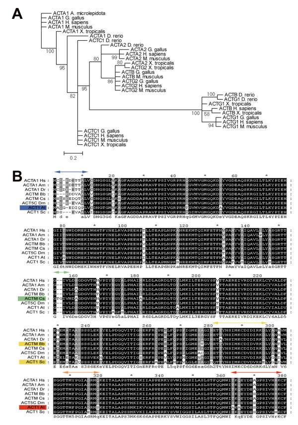
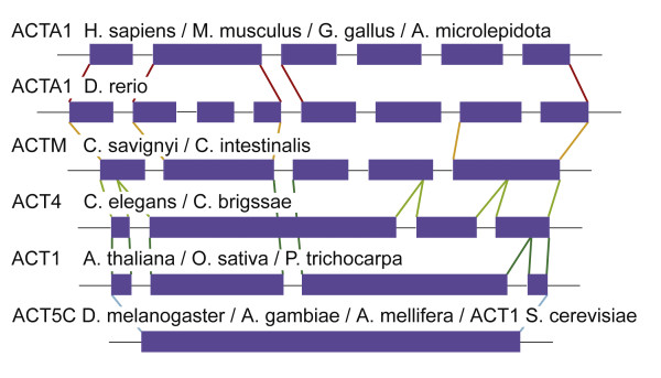
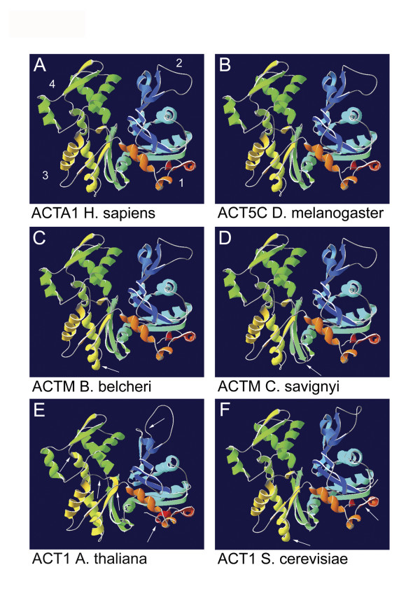
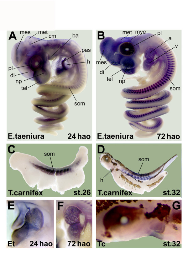
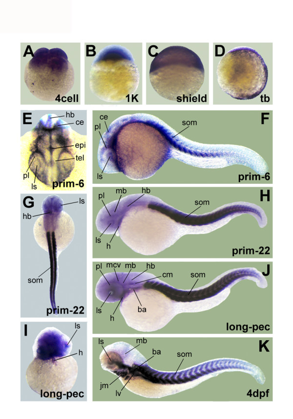
Results: We present here a model for the evolution of the genomic organization of alpha-skeletal actin and by molecular modeling, illustrate the structural differences of actin proteins of different phyla. We further describe and compare alpha-skeletal actin expression in two developmental stages of five vertebrate species (mouse, chicken, snake, salamander and fish). Our findings confirm that alpha-skeletal actin is expressed in skeletal muscle and in the heart of all five species. In addition, we identify many novel non-muscular expression domains including several in the central nervous system.

Conclusion: Our results show that the high sequence homology of alpha-skeletal actins is reflected by similarities of their 3 dimensional protein structures, as well as by conserved gene expression patterns during vertebrate development. Nonetheless, we find here important differences in 3D structures, in gene architectures and identify novel expression domains for this structural and functional important gene.


Figures

Figure 1
Figure 1
Phylogenetic tree and amino acid alignment. (A) Shown is the phylogenetic analysis of known actin isoforms from selected vertebrates numbers give the percentages for the Bayesian posterior probability. The accession numbers of the sequences used in this analysis are given in Additional file 1 and additional phylogenetic trees are given in Additional file 3. (B) The alignment of the amino acid sequences of the selected actins of human (Hs), snake (Am), zebrafish (Dr), branchiostoma (Bb), urochordate (Cs), fly (Dm), plant (At) and yeast (Sc) is showing the strong conservation of the sequence. The darker shading is used to indicate the more conserved locations. The colored arrows highlight the main sites which show a difference in the 3D protein model (Figure 3). The corresponding coloring of the protein and species name highlights which species show this difference in our 3D protein models.
Figure 2
Figure 2
Comparison of the genomic architecture. A comparison of the genomic architecture, showing the exons (on scale) and the introns (not on scale). Colored lines were added to indicate the boundaries of the exons. The asterisk marks the intron that has been conserved from plants to higher chordates, but has been lost in insects and nematodes. We have identified the intron-exon boundaries of the alpha-skeletal actin genes in several vertebrates (Homo sapiens, Mus musculus, Gallus gallus, Atractaspis microlepidota, Xenopus tropicalis, Danio rerio), chordates (Ciona savignyi, Ciona intestinalis), nematodes (Caenorhabditis elegans, Caenorhabditis briggsae), insects (Drosophila melanogaster, Drosophila pseudoobscura, Anopheles gambiae, Aedes aegypti, Culex pipiens, Apis mellifera), plants (Arabidopsis thaliana, Oryza sativa, Populus trichocarpa) and yeast (Saccharomyces cerevisia). All insects show a single exon and all vertebrates except the zebrafish (Danio rerio) show the same exon/intron boundaries as depicted for humans.
Figure 3
Figure 3
3D protein models. 3D protein models of human alpha-skeletal actin (A) and corresponding actins from fly (B), urochordate (C), branchiostoma (D), plant (E) and yeast (F). The secondary structures at the amino terminus of the protein are indicated by dark blue shading and the secondary structures at the C-terminus are colored red. The numbers indicate the subunits of the protein. White arrows point out the main differences between the models. The sites of these differences are highlighted with corresponding coloring in Figure 1.
Figure 4
Figure 4
Expression of alpha-skeletal actin in mouse and chicken embryos. Results of the in situ hybridization on embryos of mouse, stage E10.5 (A) en stage E11.5 (B). Details of the atrium are shown for the early stage embryo (E) and the later stage embryos (F). The same experiment has been conducted on chicken embryos of stage HH20 (C) and HH25 (D). Again details of the atrium are shown for both stages (G, H). Structures with staining are labeled: atrium (a) branchial arches (ba), diencephalon (di), distal progress zone (dpz), fore limb bud (flb), heart (h), hind limb bud (hlb), hyoid arch (hy), lens (ls), liver (lv), mandible (m), mandibular arch (md), maxillary arch (mx), mesencephalon (ms), metencephalon (met), myelencephalon (mye), nasal pituitary (np), otic vesicle (ov), somite (som), telencephalon (tel), ventricle (v).
Figure 5
Figure 5
Expression of alpha-skeletal actin in snake and salamander embryos. Results of the in situ hybridization on snake embryos 24 hours after oviposition (hao) (A) and 72 hao (B). Detailed pictures of the heart are shown for both stages (E, F). Two salamander embryos were also used for an in situ hybridization experiment, one staged as st.26 (C) and the older one staged as st.32 (D). A detail of the head and heart region is shown for the oldest embryo (G). The same abbreviations were used for the labeling in Figure 4. Three additional structures were labeled: cephalic musculature (cm), primary atrial septum (pas) and proliferative cell layer (pl).
Figure 6
Figure 6
Expression of alpha-skeletal actin in zebrafish embryos. Results for whole mount in situ hybridization of zebrafish embryos at several stages of development: 4cell (A), 1 K (B), shield (C), tail bud (D), prim-6 (F), prim-22 (H), long-pec (J) and 4 dpf (K). Detailed pictures of dorsal view of the head in prim-6 (E), a dorsal overview picture in prim-22 (G) and a frontal view of the head in long-pec (I) are added. The same abbreviations were used for the labeling as in Figure 4 and Figure 5. Six additional structures were labeled: cerebellum (ce), epiphysis (epi), hindbrain (hb), jaw musculature (jm), midcerebral vein (mcv) and midbrain (mb).

References

    1. Ankenbauer T, Kleinschmidt JA, Walsh MJ, Weiner OH, Franke WW. Identification of a widespread nuclear actin binding protein. Nature. 1989;8(6251):822–825. doi: 10.1038/342822a0. - DOI - PubMed
    1. Tripathi SC. A possible role of actin in the mechanical control of the cell cycle. Biology of the Cell. 1989;8(3):351–353. doi: 10.1016/0248-4900(89)90073-7. - DOI - PubMed
    1. St-Onge D, Gicquaud C. Research on the mechanism of interaction between actin and membrane lipids. Biochemical and Biophysical Research Communications. 1990;8(1):40–47. doi: 10.1016/0006-291X(90)91727-A. - DOI - PubMed
    1. Tranter M, Sugrue S, Schwartz M. Evidence for a direct, nucleotide-sensitive interaction between actin and liver cell membranes. J Cell Biol. 1989;8(6):2833–2840. doi: 10.1083/jcb.109.6.2833. - DOI - PMC - PubMed
    1. Cheng H, Bjerknes M. Asymmetric distribution of actin mRNA and cytoskeletal pattern generation in polarized epithelial cells. Journal of Molecular Biology. 1989;8(3):541–549. doi: 10.1016/0022-2836(89)90130-7. - DOI - PubMed

LinkOut - more resources