Review
. 2008 May;28(3):266-78.
doi: 10.1016/j.semnephrol.2008.03.010.

Bypassing vasopressin receptor signaling pathways in nephrogenic diabetes insipidus

Affiliations
Review

Bypassing vasopressin receptor signaling pathways in nephrogenic diabetes insipidus

Richard Bouley et al. Semin Nephrol. 2008 May.

Abstract

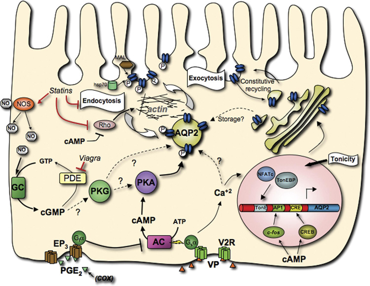
Water reabsorption in the kidney represents a critical physiological event in the maintenance of body water homeostasis. This highly regulated process relies largely on vasopressin (VP) action and on the VP-sensitive water channel (AQP2) that is expressed in principal cells of the kidney collecting duct. Defects in the VP signaling pathway and/or in AQP2 cell surface expression can lead to an inappropriate reduction in renal water reabsorption and the development of nephrogenic diabetes insipidus, a disease characterized by polyuria and polydipsia. This review focuses on the major regulatory steps that are involved in AQP2 trafficking and function. Specifically, we begin with a discussion on VP-receptor-independent mechanisms of AQP2 trafficking, with special emphasis on the nitric oxide-cyclic guanosine monophosphate signaling pathway, followed by a review of the mechanisms that govern AQP2 endocytosis and exocytosis. We then discuss emerging data illustrating roles played by the actin cytoskeleton on AQP2 trafficking, and lastly we consider elements that affect AQP2 protein expression in cells. Recent advances in each topic are summarized and are presented in the context of their potential to serve as a basis for the development of novel therapies that may ultimately improve life quality of nephrogenic diabetes insipidus patients.


Figures

Figure 1
Figure 1. Schematic representation of AQP2 trafficking in principal cells
This model shows the interactions between the components of some of the major pathways that affect AQP2, and summarizes most of the points outlined in this review. The canonical V2R signaling pathway is depicted, with VP stimulation of V2R leading to the phosphorylation of AQP2 by PKA and subsequently altering the balance between exocytosis and endocytosis, leading to AQP2 accumulation at the apical plasma membrane. Also shown are the NO-cGMP pathway and the PGE2 receptor EP3 that can also positively and negatively modulate AQP2 trafficking respectively, as well as factors that affect AQP2 abundance and the elements and transcription factors that mediate this regulation. (activator protein-1 element (AP1), adenylate cyclase (AC), aquaporin-2 (AQP2), cAMP response element (CRE), CRE binding protein (CREB), cyclooxygenase (COX), G-protein i α subunit (Giα), G-protein s α subunit (Gsα), guanylate cyclase (GC), heat-shock protein of 70kDa (hsp70), myelin and lymphocyte-associated protein (MAL), nitric oxide (NO), nitric oxide synthase (NOS), nuclear factor of activated T-cells c (NFATc), phosphodiesterase (PDE), prostaglandin E2 (PGE2), prostaglandin receptor (EP3), protein kinase G (PKG), protein kinse A (PKA), Rho family small GTPase (Rho), tonicity response element (TonE), TonE binding protein (TonEBP), vasopressin (VP), vasopressin receptor type-2 (V2R))
Figure 2
Figure 2. Western blot detection of AQP2 in plasma membrane-enriched fractions from LLC-PK1 cells expressing c-myc-tagged AQP2 (A). Indirect immunofluorescence microscopy of tissue slices showing AQP2 redistribution in the inner stripe (outer medulla) of collecting duct principal cells in response to PDE V inhibition (B)
In panel A, cells were incubated 45 min in the presence of the selective PDE5 inhibitor (sildenafil) or with the non-selective PDE inhibitor (IBMX) at a 0.1, 1 or 10 fold higher dose than that corresponding to the EC50 of either chemical agent. A plasma membrane fraction was isolated from the cells and probed with anti-AQP2 antibodies. The same plot was reprobed with an anti-pan-actin monoclonal antibody as a loading control. Both sildenafil and IBMX induce the appearance of AQP2 in the plasma membrane fraction of the cells in a does-dependent manner. In panel B kidney slices from a Sprague-Dawley rat were incubated for 10 min with (Deamino-Cys1, D-arg8)VP (DDAVP, 10 nM) or 45 min with sildenafil (0.5 µM) before fixation by immersion, sectioning and immunostaining for AQP2. Panel (A) shows a diffuse intracellular distribution of AQP2 in a control medullary collecting duct, whereas apical membrane accumulation (arrows) is induced in tissues treated with DDAVP (B) or sildenafil (C). Bar = 25 µm
Figure 3
Figure 3. AQP2 membrane accumulation can be induced by inhibiting endocytosis
Control LLC-PK1 cells expressing AQP2 displayed baseline perinuclear AQP2 staining (A), whereas cells exposed to vasopressin (VP) showed strong AQP2 expression at the plasma membrane (B). Endocytosis was blocked in LLC-PK1 cells by methyl-β-cyclodextrin (mβCD) treatment (C), expressing a dominant interfering dynamin mutant (dynamin 2 DK44A) (D) or an ATPase deficient hsc70 mutant (T204V) (E). All three approaches to reduce endocytosis resulted in a dramatic increase of AQP2 expression at the plasma membrane. Immunostaining was performed using an anti-c-myc antibody to detect the c-myc tag of AQP2 in stably transfected LLC-PK1 cells. Bar = 20 µm.
Figure 4
Figure 4. VP/FK treatment increases exocytosis in AQP2-expressing cells, but not in control cells
LLC-PK1 cells expressing AQP2 were transfected with a vector encoding a soluble, secreted form of yellow fluorescence protein, YFP (kindly provided by Jennifer Lippincott-Schwartz, NIH). The amount of ssYFP produced in LLC-ssYFP (which express YFP but not AQP2) and LLC-AQP2-ssYFP cells (which express AQP2 and YFP) and secreted in the extracellular medium after 15 min was measured by fluorimetry, and is similar between both cell lines under baseline conditions (bars 1 and 3 from left to right). When VP/FK is applied, AQP2-expressing cells show a large increase in ssYFP secretion within the first 15 min of stimulation, as compared to control cells (bars 2 and 4, respectively). These results are consistent with a large burst of exocytosis of AQP2-containing vesicles in response to VP/FK stimulation. Values were calculated as the relative increase from the 0 min baseline control and are expressed in relative fluorescence units (RFU). Each bar represents the average of 5 independent experiments performed in triplicate.

References

    1. Deen PM. Mouse models for congenital nephrogenic diabetes insipidus: what can we learn from them? Nephrol Dial Transplant. 2007;22:1023–1026. - PubMed
    1. Wade JB, Stetson DL, Lewis SA. ADH action: evidence for a membrane shuttle mechanism. Ann NY Acad Sci. 1981;372:107–117. - PubMed
    1. Kamsteeg EJ, Heijnen I, van Os CH, et al. The subcellular localization of an aquaporin-2 tetramer depends on the stoichiometry of phosphorylated and nonphosphorylated monomers. J Cell Biol. 2000;151:919–930. - PMC - PubMed
    1. Yip KP. Coupling of vasopressin-induced intracellular Ca2+ mobilization and apical exocytosis in perfused rat kidney collecting duct. J. Physiol. 2002;538:891–899. - PMC - PubMed
    1. Nickols HH, Shah VN, Chazin WJ, et al. Calmodulin interacts with the V2 vasopressin receptor: elimination of binding to the C terminus also eliminates arginine vasopressin-stimulated elevation of intracellular calcium. J Biol Chem. 2004;279:46969–46980. - PubMed

Publication types