Review
. 2008 Oct 1;80(1):9-19.
doi: 10.1093/cvr/cvn133. Epub 2008 Jun 2.

Remodelling of gap junctions and connexin expression in diseased myocardium

Affiliations
Review

Remodelling of gap junctions and connexin expression in diseased myocardium

Nicholas J Severs et al. Cardiovasc Res. .

Abstract

Gap junctions form the cell-to-cell pathways for propagation of the precisely orchestrated patterns of current flow that govern the regular rhythm of the healthy heart. As in most tissues and organs, multiple connexin types are expressed in the heart: connexin43 (Cx43), Cx40 and Cx45 are found in distinctive combinations and relative quantities in different, functionally-specialized subsets of cardiac myocyte. Mutations in genes that encode connexins have only rarely been identified as being a cause of human cardiac disease, but remodelling of connexin expression and gap junction organization are well documented in acquired adult heart disease, notably ischaemic heart disease and heart failure. Remodelling may take the form of alterations in (i) the distribution of gap junctions and (ii) the amount and type of connexins expressed. Heterogeneous reduction in Cx43 expression and disordering in gap junction distribution feature in human ventricular disease and correlate with electrophysiologically identified arrhythmic changes and contractile dysfunction in animal models. Disease-related alterations in Cx45 and Cx40 expression have also been reported, and some of the functional implications of these are beginning to emerge. Apart from ventricular disease, various features of gap junction organization and connexin expression have been implicated in the initiation and persistence of the most common form of atrial arrhythmia, atrial fibrillation, though the disparate findings in this area remain to be clarified. Other major tasks ahead focus on the Purkinje/working ventricular myocyte interface and its role in normal and abnormal impulse propagation, connexin-interacting proteins and their regulatory functions, and on defining the precise functional properties conferred by the distinctive connexin co-expression patterns of different myocyte types in health and disease.


Figures

Figure 1
Figure 1
Summary of the typical connexin expression patterns of the mammalian heart.
Figure 2
Figure 2
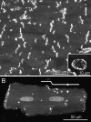
Characteristic distribution pattern of Cx43 gap junctions in ventricular myocardium. (A) Longitudinal section from rat left ventricle. The Cx43 gap junctions appear in rows, corresponding to edge-on viewed intercalated discs. Inset shows a single intercalated disc viewed face-on from transversely sectioned human myocardium. Note larger gap junctions at the periphery of the disc. (B) The presence of multiple discs of different size is best appreciated in views of isolated myocytes (in this example, from rat). The steps of the disc are indicated by the white line. Note that some apparently isolated gap junctions at the lateral surface (indicated by spot on the line) can be considered as components of extended intercalated discs.
Figure 3
Figure 3
Organization of junctions at the intercalated disc, as seen by thin-section electron microscopy. In low magnification view (A), the step-like features of the discs are clearly seen (right end of cell and arrowheads). If we take, at higher magnification, an area from within the disc like that enclosed by the box, the three junction types are visible (B). Fasciae adherentes occupy electron-dense vertical plicate zones of the disc; gap junctions and desmosomes mainly the lateral-facing zones. In this example, the ends of the gap junction contact a fascia adherens (arrows), though in many instances non-junctional intercalated disc plasma membrane separates the junctions from one another [(A) from Severs, N.J. et al J Ultrastruct Res 1982;81:222–239; reprinted with permission from Elsevier; (B) from Severs, N.J. BioEssays 2000;22:188–199].
Figure 4
Figure 4
Variations in the pattern of organization of gap junctions in atrial myocardium, as seen by Cx43 labelling in dissociated groups of rat atrial myocytes. While clusters of gap junctions in classic step-like and straight-end intercalated disc configurations are present [lines, left side of cells in (A) and (B)], the junctions often appear spread laterally (asterisks). Gap junction distribution can thus range from largely end-to-end (C) to predominantly side-to-side (D).
Figure 5
Figure 5
The Purkinje/working ventricular myocyte interface. (A) Cellular architecture. Purkinje myocytes link to underlying flattened transitional cells, which in turn link to the working ventricular myocardium. (B) immunoconfocal view of Purkinje myocyte/working ventricular myocyte interface from mouse myocardium showing transversely sectioned Purkinje myocytes (P) above a longitudinally sectioned transitional cell (T). The working ventricular myocyardium (WM) lies below. Connexin immunolabelling shows that whereas Cx40 (C) is abundant in the Purkinje myocytes and transitional cells, Cx45 (D) is seen in both these cell types and in the most superficial working ventricular myocytes [asterisk, (B)].
Figure 6
Figure 6
(A) Cx43 is down-regulated and ZO-1 up-regulated in end-stage heart failure, giving a significant negative correlation (P = 0.0029; r2 = 0.51). (B) This is associated with an increased interaction of Cx43 with ZO-1 in both non-junctional and gap-junctional fractions as determined by co-immunoprecipitation (The non-junctional fraction is Triton X-100-soluble and represents Cx43 not assembled into gap junctions). DCM, dilated cardiomyopathy; ICM, ischaemic cardiomyopathy. *P = 0.05 vs. controls. From Bruce et al.

References

    1. Harris AL, Locke D. Connexins: A Guide. Secaucus: Humana Press - Springer; 2008.
    1. Severs NJ, Coppen SR, Dupont E, Yeh HI, Ko YS, Matsushita T. Gap junction alterations in human cardiac disease. Cardiovasc Res. 2004;62:368–377. - PubMed
    1. Severs NJ, Dupont E, Thomas N, Kaba R, Rothery S, Jain R, et al. Alterations in cardiac connexin expression in cardiomyopathies. Adv Cardiol. 2006;42:228–242. - PubMed
    1. Sohl G, Willecke K. Gap junctions and the connexin protein family. Cardiovasc Res. 2004;62:228–232. - PubMed
    1. Harris AL. Emerging issues of connexin channels: biophysics fills the gap. Q Rev Biophys. 2001;34:325–472. - PubMed

Publication types