. 2008 Sep;28(17):5251-64.
doi: 10.1128/MCB.00048-08. Epub 2008 Jun 2.

Distinct functions of POT1 at telomeres

Affiliations

Distinct functions of POT1 at telomeres

Katharine S Barrientos et al. Mol Cell Biol. 2008 Sep.

Abstract

The mammalian protein POT1 binds to telomeric single-stranded DNA (ssDNA), protecting chromosome ends from being detected as sites of DNA damage. POT1 is composed of an N-terminal ssDNA-binding domain and a C-terminal protein interaction domain. With regard to the latter, POT1 heterodimerizes with the protein TPP1 to foster binding to telomeric ssDNA in vitro and binds the telomeric double-stranded-DNA-binding protein TRF2. We sought to determine which of these functions-ssDNA, TPP1, or TRF2 binding-was required to protect chromosome ends from being detected as DNA damage. Using separation-of-function POT1 mutants deficient in one of these three activities, we found that binding to TRF2 is dispensable for protecting telomeres but fosters robust loading of POT1 onto telomeric chromatin. Furthermore, we found that the telomeric ssDNA-binding activity and binding to TPP1 are required in cis for POT1 to protect telomeres. Mechanistically, binding of POT1 to telomeric ssDNA and association with TPP1 inhibit the localization of RPA, which can function as a DNA damage sensor, to telomeres.


Figures

FIG. 1.
FIG. 1.
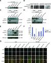
POT1 truncation mutants. (A) Diagrams of indicated POT1 proteins. OB1 and OB2, oligonucleotide/oligosaccharide DNA-binding domains; FLAG, FLAG epitope tag. Numbers give amino acid numbers. (B) In vitro immunoprecipitation of the indicated 35S-labeled F-POT1 proteins with a 32P-labeled telomeric ssDNA oligonucleotide. (C and D) Immunoprecipitation (IP) of GFP-tagged TPP1 (GFP-TPP1) (C) or myc-tagged TIN2 (myc-TIN2) (D) followed by immunoblotting (IB) to detect whether the indicated F-POT1 proteins coimmunoprecipitated with GFP-TPP1 (C) or myc-TIN2 (D) in 293T cells. Inputs were 1/10 of the total immunoprecipitation lysates. F-POT1ΔOB (C) and F-POT1 (D) are difficult to see as inputs but are clearly visible as coimmunoprecipitates with GFP-TPP1 and myc-TIN2. (E) Visualization by direct fluorescence of the indicated YFP-FLAG-tagged POT1 (YFP-F-POT1) proteins (green channel) and RFP-TRF2 proteins (red channel) in 293T cells. (F) ChIP analysis of the indicated proteins expressed in 293T cells immunoprecipitated with an anti-FLAG antibody and Southern hybridized with the indicated probes in the absence or presence of cross-linking. Total genomic DNA served as a loading control. All images are representative of duplicate experiments, except for panels C and D, which are representative of triplicate experiments.
FIG. 2.
FIG. 2.
ssDNA-binding and protein interaction activities of POT1 are required to protect telomere ends. (A) Detection of the indicated F-POT1 proteins expressed in IMR-90 cells by immunoprecipitation (IP) followed by immunoblotting (IB) with an anti-FLAG antibody. (B) Detection of endogenous POT1 mRNA, or GAPDH mRNA as a loading control, by RT-PCR in the indicated IMR-90 cells. (C and D) Detection of endogenous TRF1 (green channel) and γ-H2AX (red channel) by indirect immunofluorescence, whereby colocalization indicates TIFs, quantitated from >150 cells (C) or as representative images (D) of the indicated IMR-90 cells. Images are representative of duplicate experiments. Error bars represent standard errors. (E) Crystal violet-stained IMR-90 cells expressing the indicated transgenes 1 week after being seeded at low density.
FIG. 3.
FIG. 3.
Separation-of-function mutants of POT1. (A) Immunoprecipitation (IP) of GFP-tagged TPP1 (GFP-TPP1) followed by immunoblotting (IB) to detect whether the indicated F-POT1 proteins coimmunoprecipitated with GFP-TPP1 in 293T cells. (B) Immunoprecipitation (IP) of the indicated F-POT1 proteins followed by immunoblotting (IB) to detect whether endogenous TRF2 coimmunoprecipitated with the indicated F-POT1 proteins in 293T cells. Inputs were 1/10 of the total immunoprecipitation lysates. Images are representative of triplicate experiments.
FIG. 4.
FIG. 4.
TRF2-POT1 interaction promotes telomeric localization of POT1. (A) Visualization by direct fluorescence of the indicated YFP-F-POT1 (green channel) and RFP-TRF2 (red channel) proteins in 293T cells. (B) ChIP analysis of the indicated F-POT1 proteins expressed in 293T cells, immunoprecipitated with an anti-FLAG antibody, and Southern hybridized with the indicated probes in the absence or presence of cross-linking. Total genomic DNA served as a loading control. (C) In vitro immunoprecipitation of the indicated 35S-labeled F-POT1 proteins with a 32P-labeled telomeric ssDNA oligonucleotide. (D and E) Immunoprecipitation (IP) of myc-tagged TIN2 (myc-TIN2) followed by immunoblotting (IB) to detect whether the indicated F-POT1 proteins coimmunoprecipitated with myc-TIN2 in 293T cells with normal levels of endogenous TRF2 (D) or in 293T cells expressing TRF2 shRNA (E). (F) Immunoblot (IB) of endogenous TRF2 levels in 293T parental cells and in 293T cells transiently transfected with TRF2 shRNA. In panels D and E, inputs were 1/10 of the total immunoprecipitation lysates. All images are representative of duplicate experiments, except for panel D, which is representative of triplicate experiments.
FIG. 5.
FIG. 5.
TPP1-POT1 interaction protects telomeres from being detected as DNA damage. (A) Detection of the indicated F-POT1 proteins by immunoprecipitation (IP) followed by immunoblotting (IB) in IMR-90 cells. (B) Detection of endogenous POT1 mRNA, or GAPDH mRNA as a loading control, by RT-PCR with the indicated IMR-90 cells. (C and D) IP of GFP-tagged TPP1 (GFP-TPP1) (C) or myc-tagged TIN2 (myc-TIN2) (D) followed by IB to detect whether the indicated F-POT1 proteins coimmunoprecipitated with GFP-TPP1 or myc-TIN2 in 293T cells. (E) IP of the indicated F-POT1 proteins followed by IB to detect whether endogenous TRF2 coimmunoprecipitated with the indicated F-POT1 proteins in 293T cells. (F and G) Detection of endogenous TRF1 (green channel) and γ-H2AX (red channel) by indirect immunofluorescence, whereby colocalization indicates TIFs, quantitated from >150 cells (E) or as representative images (F) of the indicated IMR-90 cells. Error bars represent standard errors. In panels C to E, inputs were 1/10 of the total immunoprecipitation lysates. Images in panels C to G are representative of duplicate experiments.
FIG. 6.
FIG. 6.
POT1 inhibits RPA association with telomeres. Visualization by indirect immunofluorescence was performed to show endogenous TRF1 (red channel) and RPA (green channel) in IMR-90 cells expressing the indicated F-POT1 proteins or POT1 shRNA, as representative images (A) and quantitated from two experiments with >75 cells each (B). Error bars represent standard errors.
FIG. 7.
FIG. 7.
Models of distinct functions of POT1 separation-of-function mutants at telomeres. (A) Wild-type POT1 functions in a telomeric dsDNA subcomplex via direct interaction with TRF2, which enhances localization to telomeric chromatin, and in a telomeric ssDNA subcomplex via a direct interaction with TPP1 that protects telomere ends from detection as DNA damage by excluding RPA, which can act as a DNA damage sensor, from telomeric ssDNA. (B) POT1ΔTPP1 can still localize to telomeric chromatin via direct interaction with TRF2 but allows RPA access to telomeric ssDNA and thus fails to protect chromosome ends from being detected as DNA damage. (C) POT1ΔTRF2 fails to localize to telomeric chromatin but excludes RPA from telomeric ssDNA and thus protects telomeres from being detected as DNA damage. Solid lines indicate a strong interaction, while dotted lines indicate a weakened interaction.

References

    1. Armbruster, B. N., S. S. Banik, C. Guo, A. C. Smith, and C. M. Counter. 2001. N-terminal domains of the human telomerase catalytic subunit required for enzyme activity in vivo. Mol. Cell. Biol. 217775-7786. - PMC - PubMed
    1. Armbruster, B. N., K. T. Etheridge, D. Broccoli, and C. M. Counter. 2003. Putative telomere-recruiting domain in the catalytic subunit of human telomerase. Mol. Cell. Biol. 233237-3246. - PMC - PubMed
    1. Armbruster, B. N., C. M. Linardic, T. Veldman, N. P. Bansal, D. L. Downie, and C. M. Counter. 2004. Rescue of an hTERT mutant defective in telomere elongation by fusion with hPot1. Mol. Cell. Biol. 243552-3561. - PMC - PubMed
    1. Baumann, P., and T. R. Cech. 2001. Pot1, the putative telomere end-binding protein in fission yeast and humans. Science 2921171-1175. - PubMed
    1. Baumann, P., E. Podell, and T. R. Cech. 2002. Human Pot1 (protection of telomeres) protein: cytolocalization, gene structure, and alternative splicing. Mol. Cell. Biol. 228079-8087. - PMC - PubMed

Publication types