. 2008 Jun 1;22(11):1478-89.
doi: 10.1101/gad.1666208.

TopBP1 activates ATR through ATRIP and a PIKK regulatory domain

Affiliations

TopBP1 activates ATR through ATRIP and a PIKK regulatory domain

Daniel A Mordes et al. Genes Dev. .

Abstract

The ATR (ATM and Rad3-related) kinase and its regulatory partner ATRIP (ATR-interacting protein) coordinate checkpoint responses to DNA damage and replication stress. TopBP1 functions as a general activator of ATR. However, the mechanism by which TopBP1 activates ATR is unknown. Here, we show that ATRIP contains a TopBP1-interacting region that is necessary for the association of TopBP1 and ATR, for TopBP1-mediated activation of ATR, and for cells to survive and recover DNA synthesis following replication stress. We demonstrate that this region is functionally conserved in the Saccharomyces cerevisiae ATRIP ortholog Ddc2, suggesting a conserved mechanism of regulation. In addition, we identify a domain of ATR that is critical for its activation by TopBP1. Mutations of the ATR PRD (PIKK [phosphoinositide 3-kinase related kinase] Regulatory Domain) do not affect the basal kinase activity of ATR but prevent its activation. Cellular complementation experiments demonstrate that TopBP1-mediated ATR activation is required for checkpoint signaling and cellular viability. The PRDs of ATM and mTOR (mammalian target of rapamycin) were shown previously to regulate the activities of these kinases, and our data indicate that the DNA-PKcs (DNA-dependent protein kinase catalytic subunit) PRD is important for DNA-PKcs regulation. Therefore, divergent amino acid sequences within the PRD and a unique protein partner allow each of these PIK kinases to respond to distinct cellular events.


Figures
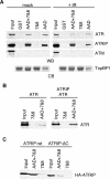
Figure 1.
Figure 1.
ATRIP promotes the association of ATR and TopBP1. (A) Nuclear extracts from 293T cells treated with 8 Gy of IR or mock-treated were incubated with equal amounts of recombinant GST-tagged fragments of TopBP1 fragments ([AAD] amino acids 978–1286 [Kumagai et al. 2006]; [7&8] BRCT repeats 7 and 8, amino acids 1182–1522; [AAD + 7&8] amino acids 978–1522) bound to glutathione beads. Proteins bound to the beads were eluted, separated by SDS-PAGE, and immunoblotted with antibodies to ATR, ATRIP, or ATM (WB). A duplicate gel was stained with Coomassie blue to verify equal amounts of GST-tagged TopBP1 proteins (CB). (B) Nuclear extracts from 293T cells transfected with a vector encoding ATR or vectors encoding ATR and ATRIP were incubated with recombinant fragments of TopBP1 bound to glutathione beads. Bound proteins were eluted, separated by SDS-PAGE, and immunoblotted with an antibody to ATR. (C) Nuclear extracts from U2OS cells stably expressing HA-tagged wild-type (wt) ATRIP or HA-tagged ATRIP lacking the C-terminal 32 amino acids (ΔC) were incubated with recombinant GST-tagged fragments of TopBP1 bound to glutathione beads. Proteins bound to the beads were eluted, separated by SDS-PAGE, and blotted with an anti-HA antibody. Input in all experiments is 5% of the extract added to the binding reaction.
Figure 2.
Figure 2.
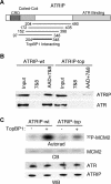
Identification of an ATRIP region necessary for TopBP1 association. (A) Schematic diagram showing the fragments of ATRIP that interacted with TopBP1 in a yeast two-hybrid assay (black lines). (CRD) Checkpoint Recruitment Domain (Ball et al. 2007). (B) Nuclear extracts from 293T cells transfected with vectors encoding ATR and wild-type ATRIP (wt) or ATR and ATRIP-top were incubated with recombinant GST-tagged fragments of TopBP1 bound to glutathione beads. Proteins bound to the beads were eluted, separated by SDS-PAGE, and blotted with antibodies to ATR or ATRIP. (C) Wild-type ATR–ATRIP (wt) or ATR–ATRIP-top complexes were isolated from transfected 293T cells and incubated with recombinant TopBP1 AAD, MCM2 substrate, and γ-32PATP. Kinase reactions were separated by SDS-PAGE, stained with Coomassie blue (CB), and exposed to film (autorad). A duplicate gel was blotted and probed with anti-ATRIP and anti-ATR antibodies (WB).
Figure 3.
Figure 3.
ATRIP association with TopBP1 is essential for cellular recovery from replication stress. (A–C) U2OS cells stably expressing siRNA-resistant wild-type ATRIP (wt), ATRIP-top, or an empty vector (vector) were transfected with siRNA targeting ATRIP to deplete endogenous ATRIP. Three days later, cells were exposed to 1 mM HU for 24 h. (A) Cells were collected immediately (0 hr) or rinsed and released into media containing 1 μg/mL nocodazole for either 8 h (8 hr) or 16 h (16 hr). Cells were fixed and stained with propidium iodine and processed for FACS analysis. (Asynch) Asynchronous cells that were not exposed to HU. (B) Immunoblot showing ATRIP levels from U2OS cells stably expressing wild-type ATRIP (wt), ATRIP-top (top), or empty vector (vt). (C) Twenty-four hours after release from HU, cellular viability was measured using a colorimetric assay. Viability was normalized to cells expressing exogenous wild-type ATRIP. Error bars indicate standard error, n = 6. (D) Three days after siRNA transfection, cells were treated with 4 Gy of IR, and 1 μg/mL nocodazole was added to the media. Sixteen hours later, the percentage of mitotic cells were determined by propidium iodine and antiphosphohistone H3 staining followed by flow cytometry.
Figure 4.
Figure 4.
An S. cerevisiae ddc2-top mutant is defective in checkpoint signaling. (A) Secondary structure prediction of the TopBP1-interacting region of ATRIP and the equivalent region of S. cerevisiae Ddc2. Hashed boxes denote the C-terminal ends of the coiled-coil domains. Solid boxes indicate predicted α-helices. The asterisk denotes the location of the ATRIP-top mutation or the ddc2-top (LLLR257AAAA) mutation. The adjacent dots in Ddc2 denote the location of the A1 (LLED274AAAA) and A2 (LIKE281AAAA) mutations. (B) Immunoblot showing Ddc2 levels in yeast strains expressing Ddc2 mutants, wild-type Ddc2, or empty vector. (C) Serial dilutions of the indicated yeast strains grown on YPD with no drug (mock), 150 mM HU (+HU), or 0.008% MMS (+MMS). (D) Exponentially growing yeast were treated with no drug (mock), 150 mM HU (+HU), or 0.015% MMS (+MMS) for 90 min. Extracts were prepared, separated by SDS-PAGE, and immunoblotted with an antibody against Rad53. The top bands are phosphorylated forms of Rad53.
Figure 5.
Figure 5.
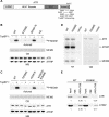
An ATR regulatory region between the kinase and FATC domains is critical for TopBP1-dependent activation of ATR in vitro. (A) Schematic diagram showing the domains of ATR and the ATR fragments that interact with TopBP1 in a yeast two-hybrid assay (black lines). (B) Wild-type ATR (WT), ATR K2589E, ATR Δ2569–2576, or ATR kinase dead (KD) proteins complexed with wild-type ATRIP were isolated from transfected 293T cells and incubated with MCM2 substrate, γ-32P-ATP, and recombinant TopBP1 AAD where indicated. Kinase reactions were separated by SDS-PAGE, stained with Coomassie blue (CB), and exposed to film (autorad). A duplicate gel was immunoblotted with anti-ATRIP and anti-ATR antibodies (WB). (C) Kinase reactions to measure TopBP1-dependent activation of wild-type ATR (WT), ATR K2589E, ATR K2587E, or ATR K2587E/K2589E were performed as in B. (D) To measure basal kinase activity plasmids encoding ATRIP and Flag-tagged wild-type ATR (WT), ATR K2589E, or empty vector were expressed in 293T cells and immunoprecipitated with anti-Flag antibodies. Complexes were incubated with MCM2 substrate and γ-32PATP. Kinase reactions were separated by SDS-PAGE, stained with Coomassie blue (CB), and exposed to film (autorad). (E) Nuclear extracts from 293T cells transfected with vectors encoding wild-type ATR (wt) and ATRIP or ATR K2589E and ATRIP were incubated with recombinant fragments of TopBP1 bound to glutathione beads. Bound proteins were eluted, separated by SDS-PAGE, and immunoblotted with ATR or ATRIP antibodies. Input equals 5% of the extract used in the binding reactions. Quantification of the immunoblot signal normalized to the input is shown.
Figure 6.
Figure 6.
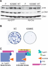
ATR K2589E does not support checkpoint signaling. (A) ATRflox/− cell lines were created that contained an inducible form of either ATR K2589E or ATR wild-type (WT). P1 and P2 denote ATRflox/− parental cell lines lacking any exogenous ATR. The cells were induced to express exogenous ATR with tetracycline and were treated with adenovirus encoding the Cre recombinase (Ad-Cre) to delete the endogenous ATR or adenovirus expressing GFP (Ad-GFP) as a control. Four days after infection, cells were treated with 1 mM HU for 6 h. Cell lysates were separated by SDS-PAGE and blotted with the indicated antibodies to ATR, Chk1, or phosphorylated Chk1. (B) Equal numbers of Cre-infected ATRflox/− cells expressing wild-type ATR or ATR K2589E were plated. Seventeen days after plating, the surviving colonies were stained with methylene blue. (C) Schematic model for ATR activation. In the absence of TopBP1, ATR exhibits a low basal kinase activity. In response to genotoxic stress, ATRIP recruits ATR to sites of DNA damage. Loading of the Rad9–Hus1–Rad1 complex allows Rad9 to recruit TopBP1. TopBP1 makes contact with both ATRIP and the ATR PRD. The interaction between TopBP1 and ATR–ATRIP greatly stimulates ATR kinase activity, perhaps due to a conformational change in the ATR kinase domain that facilitates the ability of ATR to interact with its substrates.

Comment in

References

    1. Abraham R.T. PI 3-kinase related kinases: ‘Big’ players in stress-induced signaling pathways. DNA Repair (Amst.) 2004;3:883–887. - PubMed
    1. Ball H.L., Cortez D. ATRIP oligomerization is required for ATR-dependent checkpoint signaling. J. Biol. Chem. 2005;280:31390–31396. - PMC - PubMed
    1. Ball H.L., Myers J.S., Cortez D. ATRIP binding to replication protein A-single-stranded DNA promotes ATR–ATRIP localization but is dispensable for Chk1 phosphorylation. Mol. Biol. Cell. 2005;16:2372–2381. - PMC - PubMed
    1. Ball H.L., Ehrhardt M.R., Mordes D.A., Glick G.G., Chazin W.J., Cortez D. Function of a conserved checkpoint recruitment domain in ATRIP proteins. Mol. Cell. Biol. 2007;27:3367–3377. - PMC - PubMed
    1. Banin S., Moyal L., Shieh S., Taya Y., Anderson C.W., Chessa L., Smorodinsky N.I., Prives C., Reiss Y., Shiloh Y., et al. Enhanced phosphorylation of p53 by ATM in response to DNA damage. Science. 1998;281:1674–1677. - PubMed

Publication types