. 2008 Jun 1;22(11):1490-500.
doi: 10.1101/gad.1662308.

Akt-dependent regulation of NF-{kappa}B is controlled by mTOR and Raptor in association with IKK

Affiliations

Akt-dependent regulation of NF-{kappa}B is controlled by mTOR and Raptor in association with IKK

Han C Dan et al. Genes Dev. .

Abstract

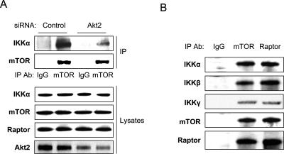
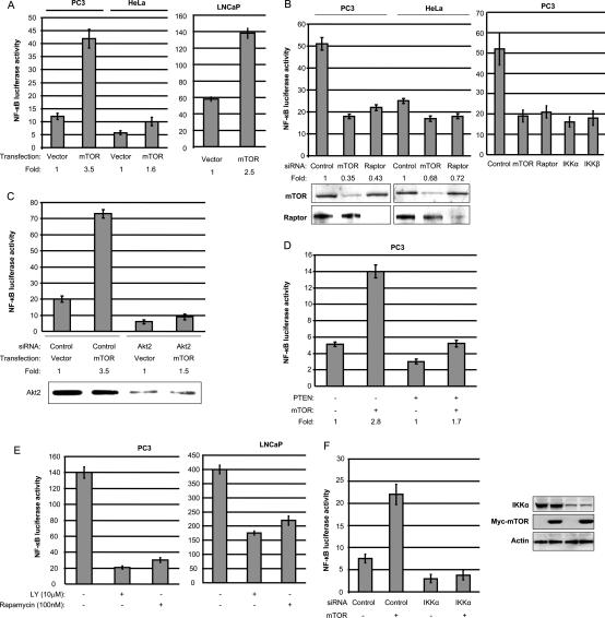
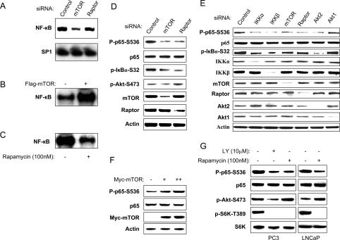
While NF-kappaB is considered to play key roles in the development and progression of many cancers, the mechanisms whereby this transcription factor is activated in cancer are poorly understood. A key oncoprotein in a variety of cancers is the serine-threonine kinase Akt, which can be activated by mutations in PI3K, by loss of expression/activity of PTEN, or through signaling induced by growth factors and their receptors. A key effector of Akt-induced signaling is the regulatory protein mTOR (mammalian target of rapamycin). We show here that mTOR downstream from Akt controls NF-kappaB activity in PTEN-null/inactive prostate cancer cells via interaction with and stimulation of IKK. The mTOR-associated protein Raptor is required for the ability of Akt to induce NF-kappaB activity. Correspondingly, the mTOR inhibitor rapamycin is shown to suppress IKK activity in PTEN-deficient prostate cancer cells through a mechanism that may involve dissociation of Raptor from mTOR. The results provide insight into the effects of Akt/mTOR-dependent signaling on gene expression and into the therapeutic action of rapamycin.


Figures

Figure 1.
Figure 1.
Akt promotes mTOR association with IKKα. (A) PC3 cells were transfected with siRNA control or siRNA to Akt2 as indicated. The cells were lysed 48 h after transfection and the lysates were immunoprecipitated with anti-mTOR, electrophoresed on an SDS gel, and blotted with mTOR and IKKα antibodies, respectively (see Dan et al. 2007). Lysates were probed with these antibodies plus anti-Raptor and anti-Akt2. (B) The lysates were immunoprecipitated with anti-mTOR, anti-Raptor, and IgG control; electrophoresed on an SDS gel; and blotted with mTOR, Raptor, IKKα, and IKKβ, and IKKγ antibodies.
Figure 2.
Figure 2.
Size fractionation demonstrates that the IKK complex is associated with mTOR–Raptor. (A) PC3 cell lysates were separated on a Superose 6 10/300 GL column. The indicated fractionations (top panel) and their mTOR immunoprecipitates (bottom panel) were analyzed by immunoblotting with the indicated antibodies. (B, top panel) PC3 cell were transfected with control siRNA and siRNA akt2, and Western blot shows the mTOR and Raptor expression. (Bottom panel) Lysates were separated on a Superose 6 10/300 GL column and the indicated fractions were analyzed by immunoblotting with the indicated antibodies.
Figure 3.
Figure 3.
mTOR/Raptor positively regulates NF-κB-dependent luciferase reporter activity in PTEN-deficient prostate cancer cells in a manner dependent on Akt. (A) PC3, LNCaP, and HeLa cells were transfected with 700 ng of mTOR expression vector or empty vector plus 200 ng of 3× κB luciferase reporter and 30 ng of pRL-SV40 (Renilla reporter control). Cells were harvested after 24 h and luciferase assays were performed (measured as relative luciferase/luminescence units). (B) PC3 and HeLa cells were transfected with the NF-κB-dependent luciferase reporter and control siRNA, or siRNA to mTOR, Raptor, IKKα, or IKKβ, as indicated. Luciferase assays were performed as in A after 24 h. Levels of luciferase are compared with the siRNA control for each cell type. Western blot shows the mTOR and Raptor protein levels. (C) PC3 cells were transfected the NF-κB-dependent luciferase reporter and either empty vector or mTOR expression vector, and with control siRNA, or siRNA to Akt2, as indicated. Luciferase assays were performed as in A after 48-h transfection of siRNA. (D) PC3 cells were cotransfected with mTOR and/or PTEN with 3× κB luciferase reporter and 30 ng of pSV40-RL as indicated. Luciferase assays were performed as in A after 24 h transfection. (E) PC3 and LNCaP cells were transfected as described above, and then treated with LY294002 (3 h) or Rapamycin (3 h) 24 h after transfection. Luciferase assays were performed as in A. SD is shown and is representative of at least three experiments. (F) PC3 cells were transfected with control siRNA, or siRNA to IKKα, as indicated. The NF-κB-dependent luciferase reporter and either empty vector or mTOR expression were transfected 24 h after siRNA transfection, and luciferase assays were performed as in A after 48-h transfection of siRNA. Immunoblotting for expressed proteins is shown. Experiments were performed in triplicate and SD is shown.
Figure 4.
Figure 4.
mTOR/Raptor is involved in control of NF-κB target gene expression in PC3 cells. PC3 cells were transfected with control siRNA, or siRNA to mTOR or Raptor, as indicated. RNA was extracted 48 h after transfection and RT–PCR (see the Materials and Methods) was performed to assess changes in mRNA levels of NF-κB target gene expression. A representative experiment from among three others is shown.
Figure 5.
Figure 5.
Akt/mTOR/Raptor pathway promotes NF-κB DNA-binding activity and RelA/p65 and IκBα phosphorylation. (A) PC3 cells were transfected with control siRNA, or siRNAs to mTOR or Raptor, as indicated. EMSAs were performed using nuclear extracts from cells lysed 48 h after transfection. A radiolabeled SP1 probe was used to normalize protein loading. (B) PC3 cells were transfected with mTOR expression vector or empty vector (control). EMSAs were performed using nuclear extracts from cells 48 h after transfection. (C) PC3 cells were treated with Rapamycin (100 nM) or DMSO for 2 h. Electrophoretic gel shift assays were performed using nuclear extracts from the cells as in A. (D,E) PC3 cells were transfected with siRNA control or other siRNAs, as indicated (IKKα, IKKβ, mTOR, Raptor, Akt1, or Akt2). The cells were lysed 48 h after transfection and the levels of IKKα, IKKβ, mTOR, Raptor, Akt1, Akt2, and β-tubulin, and of endogenous phosphorylation of P65, Akt, and IκBα were determined by immunoblotting with the indicated antibodies. (F) PC3 cells were transfected with myc-mTOR wild type or vector control as indicated. Cell lysates were generated and blotted with phospho-p65-S536, p65, S6K and myc tag, and actin antibodies, as indicated. (G) PC3 cells were treated with Rapamycin (100 nM) or DMSO for 2 h. Cell lysates were generated and blotted with the antibodies as indicated. Experiments are representative of three replicates.
Figure 6.
Figure 6.
mTOR/Raptor pathway enhances IKK kinase activity in Akt-active cells. (A) Depletion of mTOR and Raptor with siRNA decreases phosphorylation of IKKα and IKKβ in their activation loops. (B,C) Depletion of mTOR and Raptor with siRNA decreases IKK activity in PC3 cells, as measured by in vitro IKK kinase assay using GST-IκBα or GST-p65 as substrate. (D,E) PC3 cells were transfected with myc-mTOR or vector control. Endogenous IKKα, IKKβ, or IKKγ were immunoprecipitated and IKK kinase activity directed toward GST-p65 was determined in the immunoprecipitates. (F) PC3 cell lysates were separated on a Superose 6 10/300 GL column. Anti-mTOR precipitates from the indicated fractionations were incubated with GST-IκBα (1–54) and 32P-γ-ATP for kinase assay and were analyzed by immunoblotting with the indicated antibodies.
Figure 7.
Figure 7.
Rapamycin inhibits IKK activity and dissociates Raptor from the mTOR complex. (A) PC3 cells were treated with rapamycin (100 nM) or DMSO for 2 h. Cell lysates were generated and IKK activity was measured by in vitro IKK kinase assay using GST-IκBα as substrate. (B) PC3 cells were treated with rapamycin as in A. Cell lysates were immunoprecipitated with anti-mTOR and blotted with mTOR, Raptor, and IKKα antibodies, respectively.

References

    1. Agarwal A., Das K., Lerner N., Sathe S., Cicek M., Casey G., Sizemore N. The Akt/IKK pathway promotes angiogenic/metastatic gene expression in colorectal cancer by activating NF-κB and β-catenin. Oncogene. 2005;24:1021–1031. - PubMed
    1. Basseres D., Baldwin A. NF-κB and IKK pathways in oncogenic initiation and progression. Oncogene. 2006;25:6817–6830. - PubMed
    1. Bhaskar P.T., Hay N. The two TORCs and Akt. Dev. Cell. 2007;12:487–502. - PubMed
    1. Bonizzi G., Karin M. The two NF-κB activation pathways and their roles in innate and adaptive immunity. Trends Immunol. 2004;25:280–288. - PubMed
    1. Cantley L.C., Neel B.G. New insights into tumor suppression: PTEN suppresses tumor formation by restraining the PI3K/Akt pathway. Proc. Natl. Acad. Sci. 1999;96:4240–4245. - PMC - PubMed

Publication types