. 2008 Jun 2;181(5):761-75.
doi: 10.1083/jcb.200710049.

Desmin mediates TNF-alpha-induced aggregate formation and intercalated disk reorganization in heart failure

Affiliations

Desmin mediates TNF-alpha-induced aggregate formation and intercalated disk reorganization in heart failure

Panagiota Panagopoulou et al. J Cell Biol. .

Abstract

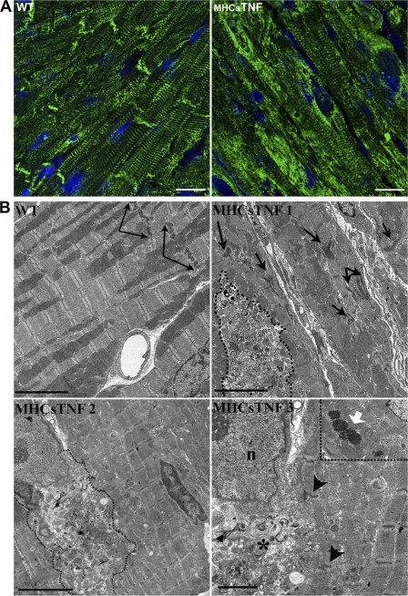
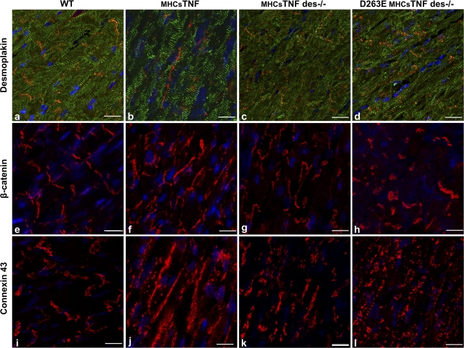
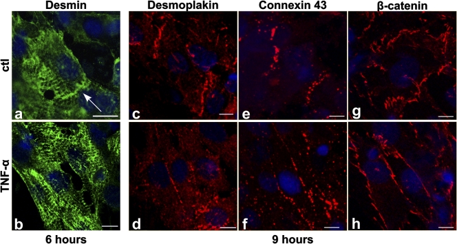
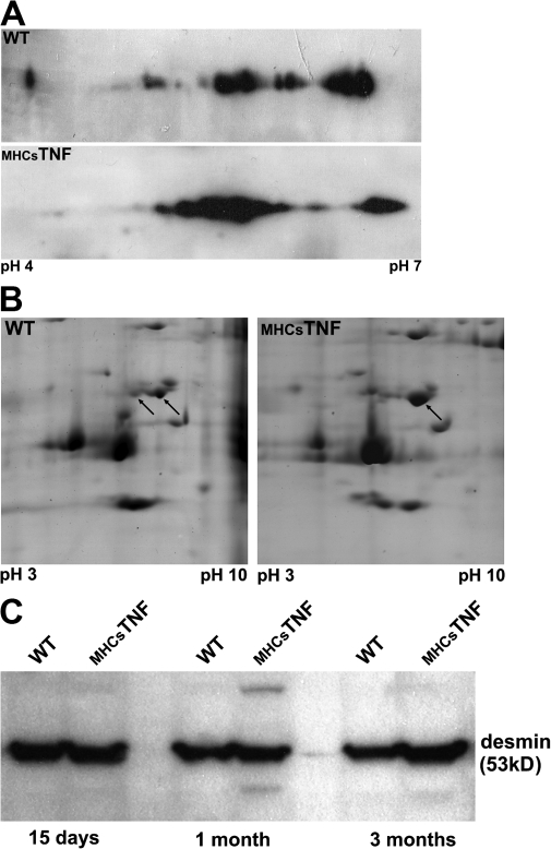
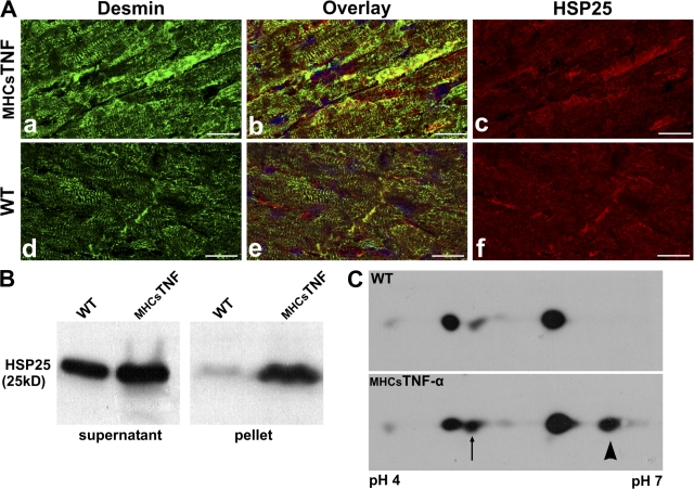
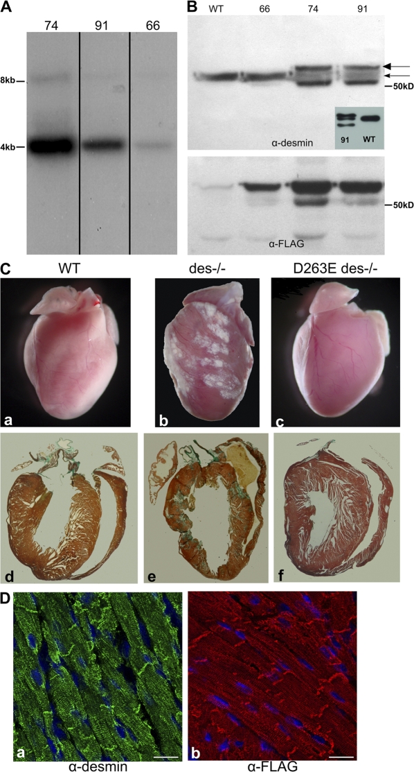
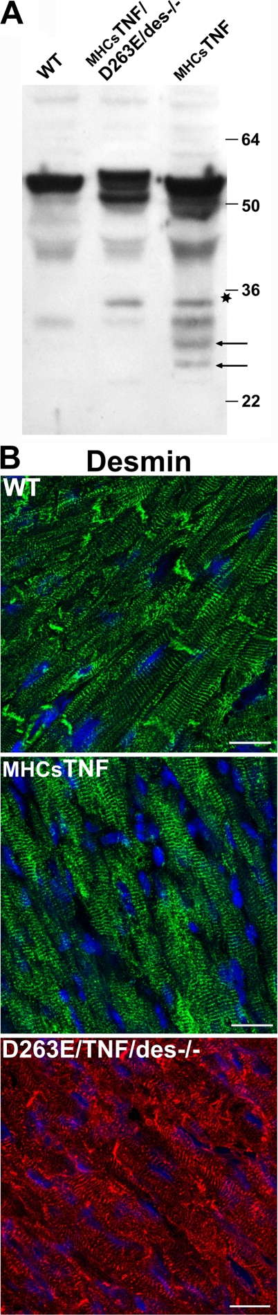
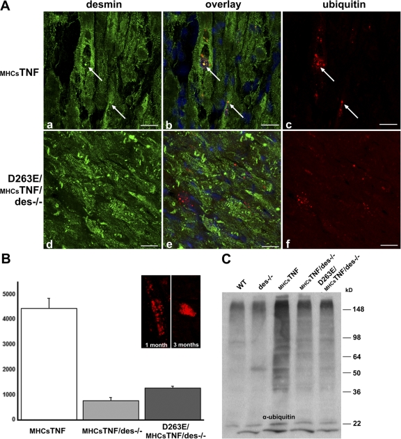
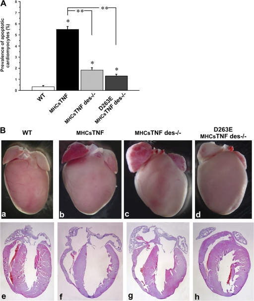
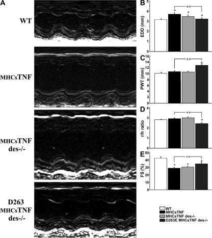
We explored the involvement of the muscle-specific intermediate filament protein desmin in the model of tumor necrosis factor alpha (TNF-alpha)-induced cardiomyopathy. We demonstrate that in mice overexpressing TNF-alpha in the heart (alpha-myosin heavy chain promoter-driven secretable TNF-alpha [MHCsTNF]), desmin is modified, loses its intercalated disk (ID) localization, and forms aggregates that colocalize with heat shock protein 25 and ubiquitin. Additionally, other ID proteins such as desmoplakin and beta-catenin show similar localization changes in a desmin-dependent fashion. To address underlying mechanisms, we examined whether desmin is a substrate for caspase-6 in vivo as well as the implications of desmin cleavage in MHCsTNF mice. We generated transgenic mice with cardiac-restricted expression of a desmin mutant (D263E) and proved that it is resistant to caspase cleavage in the MHCsTNF myocardium. The aggregates are diminished in these mice, and D263E desmin, desmoplakin, and beta-catenin largely retain their proper ID localization. Importantly, D263E desmin expression attenuated cardiomyocyte apoptosis, prevented left ventricular wall thinning, and improved the function of MHCsTNF hearts.


Figures

Figure 1.
Figure 1.
Desmin is absent from IDs and forms cytoplasmic aggregates in MHCsTNF mice. (A) Representative myocardial cryosections from 3-mo-old WT and MHCsTNF littermates were labeled for desmin and analyzed by confocal microscopy. Nuclei were stained with DAPI. (B) Electron micrographs from 3-mo-old WT and MHCsTNF mice. Arrows in WT point to IDs. Perinuclear aggregates and sarcoplasmic aggregates are displayed in TNF 2 and 1, respectively. Aggregates are outlined with dashed lines. TNF 3 is an enlargement of TNF 2 showing Z disks adjacent to an aggregate (asterisk) with material apparently merging into the aggregate (arrowheads). The white arrow in the inset of TNF 3 shows aggregates with electron-dense material. n, nucleus. Bars: (A) 20 μm; (B, top and bottom left) 5 μm; (B, bottom right) 2 μm.
Figure 2.
Figure 2.
Desmin is implicated in TNF-α–induced desmoplakin and β-catenin mislocalization. Myocardial cryosections from WT (a, e, and i), MHCsTNF (b, f, and j), MHCsTNF des−/− (c, g, and k), and D263E MHCsTNF des−/− (d, h, and l) mice at 3 mo of age were labeled for β-catenin (e–h) and connexin 43 (i–l) and were double labeled for α-actinin (green) and desmoplakin (red; a–d) followed by confocal microscopy analysis. Double labeling with α-actinin was done in all cases but is shown only in one (with desmoplakin) just to visualize the orientation of the cells; it is not shown for the rest because its high intensity masks the ID signal. Nuclei were stained with DAPI. Bars, 20 μm.
Figure 3.
Figure 3.
Addition of TNF-α to in vitro cultured cardiomyocytes induces the withdrawal of desmin, β-catenin, desmoplakin, and connexin 43 from the ID structure. Neonatal rat cardiomyocytes were incubated with TNF-α for the indicated time periods (b, d, f, and h). Cells were immunostained for desmin (a and b), desmoplakin (c and d), connexin 43 (e and f), and β-catenin (g and h) and analyzed by confocal microscopy. The arrow indicates the presence of desmin at IDs in the control culture. Nuclei were stained with DAPI. Bars, 10 μm.
Figure 4.
Figure 4.
Accumulation and modification of desmin in the MHCsTNF heart. (A) Crude extracts from hearts of 3-mo-old WT and MHCsTNF mice were electrophoretically analyzed in the first dimension by using pH 4–7 IPGphor strips and in the second dimension by 12% SDS-PAGE followed by immunoblotting using antibody for desmin. (B) Gels similar to those transferred to polyvinylidene difluoride membranes (IPG-strips pH 3–10) were counterstained with colloidal Coomassie Brilliant blue to visualize the protein loading. The arrows indicate spots identified as desmin by mass spectrophotometry. (C) Representative Western blot showing the protein expression levels of desmin at mice of different ages (15 d and 1 and 3 mo).
Figure 5.
Figure 5.
HSP25 is modified and colocalizes with desmin in subcellular aggregates of the MHCsTNF myocardium. (A) Immunofluorescent data from ventricular cryosections of 3-mo-old WT (d–f) and MHCsTNF (a–c) littermates were double labeled for desmin (a and d; green) and HSP25 (c and f; red) and analyzed by confocal microscopy. Nuclei were stained with DAPI. (B) Protein expression levels of HSP25 in 3-mo-old WT and MHCsTNF mice. (C) Western blot analysis of HSP25 from 2D gel electrophoresis of hearts from WT and MHCsTNF mice. Total protein extracts were electrophoretically analyzed in the first dimension by using pH 4–7 IPGphor strips and in the second dimension by 12% SDS-PAGE followed by immunoblotting analysis using antibody for murine HSP25. The arrow indicates the acidic isovariant that is increased in the MHCsTNF myocardium, and the arrowhead shows the alkaline isovariant that is present only in the sample from the MHCsTNF heart. Bars, 20 μm.
Figure 6.
Figure 6.
D263E transgenic desmin rescues the des−/− phenotype. Morphological and histological analysis. (A) Southern blot analysis from genomic DNA of the three different lines generated (66, 74, and 91), revealing the difference in transgene copy number. (B) Desmin protein expression in the three lines. Heart homogenates were analyzed in 10% SDS-PAGE and immunoblotted using anti-Flag and antidesmin antibodies. The thin arrow indicates the endogenous desmin, and the thick arrow shows the Flag-tagged desmin. Hearts from line 66 express mainly the endogenous desmin, but overexposure of the film shows that the mutant D263E desmin is expressed at low levels (not depicted); this is also confirmed by the α-Flag. The bottom band is most possibly a desmin degradation product that is always visible at high desmin concentrations. The consistency of this band has been verified with other desmin antibodies, which are shown in the inset. (C) Gross morphology of hearts (top) and masson-stained paraffin sections (bottom) from 3-mo-old WT (a and d), des−/− (b and e), and D263E des−/− (c and f) animals. (D) Representative myocardial cryosections from D263E des−/− mice were labeled for desmin (a) and desmin-Flag (b) and analyzed using confocal microscopy. Bars, 20 μm.
Figure 7.
Figure 7.
TNF-α–induced desmin cleavage is responsible for its relocalization away from the IDs. (A) Heart homogenates from WT, MHCsTNF, and D263E MHCsTNF des−/− mice were analyzed in 12% SDS-PAGE and immunoblotted with polyclonal antibody recognizing the entire desmin molecule. The arrows indicate the two TNF-α–induced caspase cleavage desmin fragments present only in TNF-α hearts. The asterisk shows a band that seems to be a TNF-α–related degradation product absent from WT. The other bands, which are common to the three specimens, are general desmin degradation products. (B) Representative myocardial cryosections from 3-mo-old WT, MHCsTNF, and D263E MHCsTNF des−/− mice were labeled for desmin and analyzed using confocal microscopy. Bars, 20 μm.
Figure 8.
Figure 8.
The TNF-α–induced ubiquitin aggregates are decreased and do not colocalize with desmin in D263E MHCsTNF des−/− mice. Cryosections from 3-mo-old MHCsTNF (a–c) and D263E MHCsTNF des−/− (d–f) mice were double immunolabeled for ubiquitin (c and f; red) and desmin (a and d; green). The arrows indicate ubiquitin aggregates that colocalize with desmin in MHCsTNF hearts. The field from the D263E MHCsTNF des−/− heart shown is not representative and was only selected to demonstrate the lack of colocalization with D263E desmin. (B) Quantification of the ubiquitin aggregates from 3-mo-old MHCsTNF, MHCsTNF des−/−, and D263E MHCsTNF des−/− mice. These aggregates are diffused in the cytoplasm at the age of 1 mo and, in older mice, are compacted near the nucleus, as shown in the inset. Error bars represent SEM. (C) Ubiquitin levels in WT, des−/−, MHCsTNF, MHCsTNF des−/−, and D263E MHCsTNF des−/− 1-mo-old mice. Representative Western blot of the soluble fraction of these hearts. Bars, 20 μm.
Figure 9.
Figure 9.
Characterization of MHCsTNF des−/− and D263E MHCsTNF des−/− mice. Measurement of apoptotic nuclei. (A) The percent prevalence of cardiomyocyte apoptosis was determined by in situ DNA ligation method in 3-mo-old WT, MHCsTNF, MHCsTNF des−/−, and D263E MHCsTNF des−/− animals. *, P < 0.05 compared with WT; **, P < 0.05 between MHCsTNF and MHCsTNF des−/− or D263E MHCsTNF. Error bars represent SEM. (B) Gross morphology (a–d) and hematoxylin/eosin-stained paraffin sections (e–h) from 3-mo-old mice of the aforementioned categories.
Figure 10.
Figure 10.
Effect of D263E desmin on LV remodeling. (A) Representative M-mode echocardiograms for WT, MHCsTNF, MHCsTNF des−/−, and D263E MHCsTNF des−/− mouse hearts. (B–E) Group data for LV EDD (B), PWT (C), r/h (D), and percent FS (E) of the aforementioned mice categories. *, P < 0.05 compared with WT; **, P < 0.05 between MHCsTNF and/or D263E MHCsTNF. Error bars represent SEM.

References

    1. Bajaj, G., and R.K. Sharma. 2006. TNF-alpha-mediated cardiomyocyte apoptosis involves caspase-12 and calpain. Biochem. Biophys. Res. Commun. 345:1558–1564. - PubMed
    1. Bar, H., S.V. Strelkov, G. Sjoberg, U. Aebi, and H. Herrmann. 2004. The biology of desmin filaments: how do mutations affect their structure, assembly, and organisation? J. Struct. Biol. 148:137–152. - PubMed
    1. Bence, N.F., R.M. Sampat, and R.R. Kopito. 2001. Impairment of the ubiquitin-proteasome system by protein aggregation. Science. 292:1552–1555. - PubMed
    1. Bennett, E.J., N.F. Bence, R. Jayakumar, and R.R. Kopito. 2005. Global impairment of the ubiquitin-proteasome system by nuclear or cytoplasmic protein aggregates precedes inclusion body formation. Mol. Cell. 17:351–365. - PubMed
    1. Brancolini, C., A. Sgorbissa, and C. Schneider. 1998. Proteolytic processing of the adherens junctions components beta-catenin and gamma-catenin/plakoglobin during apoptosis. Cell Death Differ. 5:1042–1050. - PubMed

Publication types