. 2008 Jun 17;117(24):3099-108.
doi: 10.1161/CIRCULATIONAHA.108.767673. Epub 2008 Jun 2.

Follistatin-like 1 is an Akt-regulated cardioprotective factor that is secreted by the heart

Affiliations

Follistatin-like 1 is an Akt-regulated cardioprotective factor that is secreted by the heart

Yuichi Oshima et al. Circulation. .

Abstract

Background: The Akt protein kinase is an important mediator of cardiac myocyte growth and survival. To identify factors with novel therapeutic applications in cardiac diseases, we focused on the identification of factors secreted from Akt1-activated cells that have cardioprotective effects through autocrine/paracrine mechanisms.

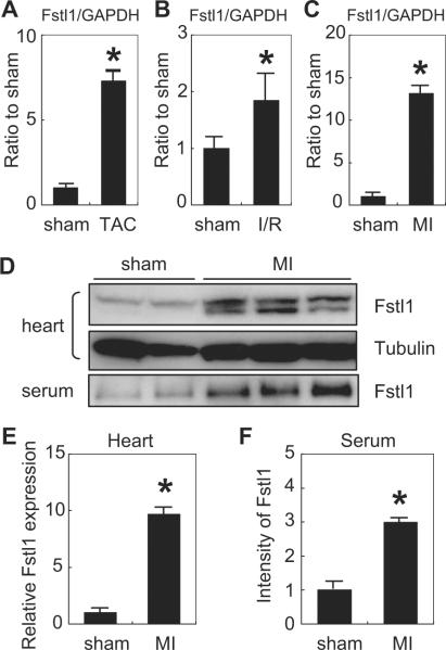
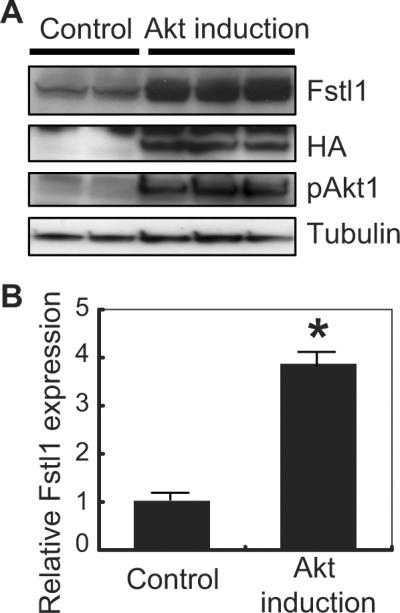
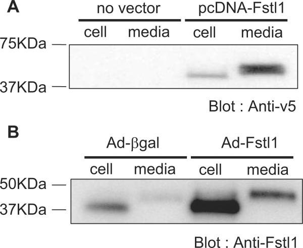
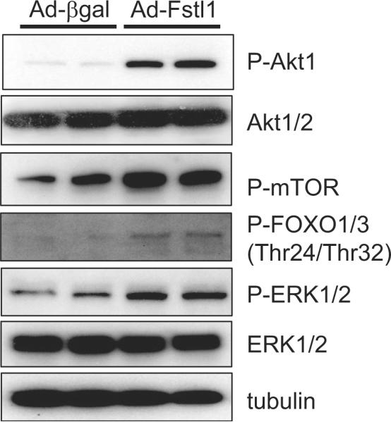
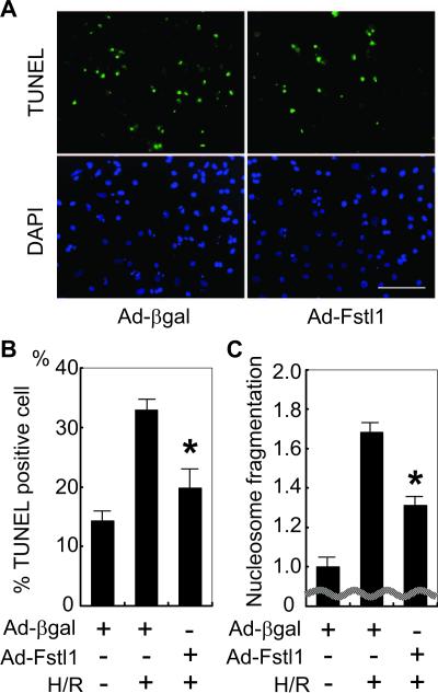
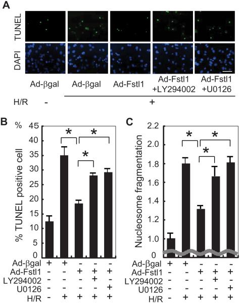
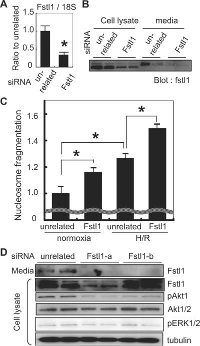
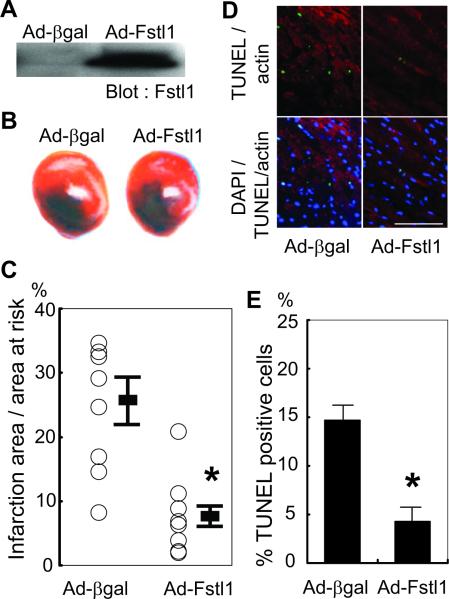
Methods and results: Using an inducible Akt1 transgenic mouse model, we have found that follistatin-like 1 (Fstl1) protein and transcript expression are increased 4.0- and 2.0-fold, respectively, by Akt activation in the heart (P<0.05). Fstl1 transcript was also upregulated in response to myocardial stresses including transverse aortic constriction, ischemia/reperfusion injury, and myocardial infarction. Adenovirus-mediated overexpression of Fstl1 protected cultured neonatal rat ventricular myocytes from hypoxia/reoxygenation-induced apoptosis (P<0.01), and this protective effect was dependent on the upregulation of both Akt and ERK activities. Conversely, knockdown of Fstl1 in cardiac myocytes decreased basal Akt signaling and increased the frequency of apoptotic death in vitro (P<0.01). The intravenous administration of an adenoviral encoding Fstl1 to mice resulted in a 66.0% reduction in myocardial infarct size after ischemia/reperfusion injury that was accompanied by a 70.9% reduction in apoptosis in the heart (P<0.01).

Conclusions: These results indicate that Fstl1 is a cardiac-secreted factor that functions as an antiapoptotic protein. Fstl1 could play a role in myocardial maintenance and repair in response to harmful stimuli.


Figures

Figure 1
Figure 1
Upregulation of Fstl1 by pathological stimuli in the heart. A to C, QRT-PCR analysis of Fstl1 transcript expression of Fstl1 mRNA after transverse aortic constriction (TAC) (A), ischemia/reperfusion (I/R) injury (B), and myocardial infarction (MI) (C) resulting from permanent LAD ligation. The data are normalized to the intensity of the GAPDH signal and are compared with Fstl1 transcript expression level in sham-operated heart. n=3 to 6. *P<0.05 vs sham. D, Western immunoblot analysis of Fstl1 expression in mouse heart lysate and serum after sham operation (left 2 lanes) and myocardial infarction (right 4 lanes). E, Quantification of Fstl1 protein expression in hearts of mice subjected to sham surgery or myocardial infarction. F, Quantification of Fstl1 protein levels in serum of mice subjected to sham surgery or myocardial infarction.
Figure 2
Figure 2
Upregulation of Fstl1 in Akt-activated heart. A, Western blots from transgenic mouse heart lysate. Transgene (myr-Akt tagged with hemagglutinin [HA]) induction was confirmed by probing with antibody against hemagglutinin (middle panel). Two weeks after myr-Akt transgene induction (lanes 3, 4, and 5), Fstl1 expression level was upregulated (top panel) compared with control mice that undergo the same doxycycline administration/withdrawal protocol (lanes 1 and 2). Each lane represents a difference transgenic or control mouse. B, Quantification of the intensity of the bands for Fstl1 normalized with that for α-tubulin. n=3 to 4. *P<0.05.
Figure 3
Figure 3
Fstl1 is secreted from cells. A, HEK293 cells were transfected with a plasmid expressing V5 epitope-tagged Fstl1 protein. Western blot analysis using anti-V5 antibody indicates that Fstl1 is present in the cell pellet and the culture media. B, NRVMs were transfected with an adenovirus expressing Fstl1 or β-galactosidase. Western blot analysis using anti-Fstl1 antibody revealed Fstl1 protein expression in the cell pellet and the culture media, and the intensity of these bands increased in cells that were transduced with Ad-Fstl1.
Figure 4
Figure 4
Activation of intracellular signaling pathways by Fstl1. After transfection of Ad-Fstl1, NRVMs were cultured for 36 hours, and cell lysates were prepared for Western blot analysis. Transduction of Ad-Fstl1 led to the activating phosphorylation of Akt1 and 2 and ERK1/2 in NRVMs. Phosphorylation of FOXO1 and 3 and mTOR, downstream targets of Akt signaling, also increased.
Figure 5
Figure 5
Fstl1 inhibits hypoxia/reoxygenationinduced apoptosis. A, Serum-deprived NRVMs underwent 12 hours of hypoxia followed by 24 hours of reoxygenation. Representative photographs of cultures stained with TUNEL (green; top panels) to identify apoptotic nuclei and DAPI (blue; bottom panels) to identify total nuclei are shown. Cultures were transfected with Ad-Fstl1 or Ad-βgal before hypoxia/reoxygenation. Scale bar=100 μm. B, Quantification of TUNEL-positive cell number. *P<0.01 compared with Ad-βgal-transfected NRVMs after hypoxia/reoxygenation (H/R). C, Nucleosome fragmentation enzyme-linked immunosorbent assay shows that transduction with Ad-Fstl1 reduced apoptosis after hypoxia/reoxygenation. *P<0.01 compared with Ad-βgal-transfected NRVMs after hypoxia/reoxygenation.
Figure 6
Figure 6
Fstl1-mediated cytoprotection is mitigated by PI3K or MEK1 inhibition. A, Representative photographs of adenovirus-transduced NRVMs stained with TUNEL (green; top panels) and DAPI (blue; bottom panels). Three hours before the induction of hypoxia, NRVMs were pretreated with a PI3K inhibitor (LY294002; 10 μmol/L), a MEK1 inhibitor (U0126; 10 μmol/L), or vehicle. Apoptotic nuclei were identified by TUNEL staining. Scale bar=50 μm. B, Quantification of TUNEL-positive cells under different culture conditions. *P<0.05. C, Nucleosome fragmentation enzyme-linked immunosorbent assay showed that Fstl1-mediated protection of myocytes from apoptosis was reversed by treatment with the PI3K inhibitor or the MEK1 inhibitor. *P<0.05. H/R indicates hypoxia/reoxygenation.
Figure 7
Figure 7
Knockdown of endogenous Fstl1 increases apoptosis in response to hypoxia/reoxygenation. A, QRT-PCR analysis indicates that transduction of siRNA decreased Fstl1 mRNA by ≈70% (*P<0.05) in cultured cardiac myocytes. B, Representative immunoblot analysis of Fstl1 protein expression in cell lysates and culture media of cardiac myocyte cultures treated with siRNA directed to Fstl1 or an unrelated sequence. C, Nucleosome fragmentation assay shows that knockdown of endogenous Fstl1 expression in serum-deprived cardiac myocyte cultures increases apoptosis in cells under normoxic conditions and in cells treated with hypoxia/reoxygenation (H/R) (*P<0.05). D, Immunoblot analysis reveals that Fstl1 ablation results in decreased Akt phosphorylation, but not that of ERK, in cultured cardiac myocytes. Two commercial sources of siRNA targeting Fstl1 were employed in these assays.
Figure 8
Figure 8
Fstl1 protects against myocardial ischemia/reperfusion injury in vivo. A, Western blot analysis of serum proteins from mice after the intravenous delivery of the indicated adenoviral vector. Sera were collected and analyzed 5 days after adenovirus delivery. Serum Fstl1 level was markedly increased by administration of Ad-Fstl1. B, Representative photographs of heart sections stained with Evans blue and 2-3-5-triphenyl tetrazolium chloride to detect the infarction zone resulting from 30 minutes of ischemia and 24 hours of reperfusion. Ischemia/reperfusion injury was performed 5 days after adenoviral injection. C, Quantification of infarction size of each experimental group (n=10 for Ad-βgal group; n=7 for Ad-Fstl1 group). *P<0.05 compared with Ad-βgal-injected group. D, Representative fluorescent images of heart sections stained with TUNEL (green), sarcomeric actin (red), and DAPI (blue). Scale bar=100 μm. E, Quantification of TUNEL-positive myocyte number showed that administration of Ad-Fstl1 decreased apoptosis (n=3 for each group). *P<0.01 compared with Ad-βgal-injected group.

References

    1. Shiojima I, Walsh K. Regulation of cardiac growth and coronary angiogenesis by the Akt/PKB signaling pathway. Genes Dev. 2006;20:3347–3365. - PubMed
    1. Fujio Y, Nguyen T, Wencker D, Kitsis RN, Walsh K. Akt promotes survival of cardiomyocytes in vitro and protects against ischemiareperfusion injury in mouse heart. Circulation. 2000;101:660–667. - PMC - PubMed
    1. Miao W, Luo Z, Kitsis RN, Walsh K. Intracoronary, adenovirus-mediated Akt gene transfer in heart limits infarct size following ischemiareperfusion injury in vivo. J Mol Cell Cardiol. 2000;32:2397–2402. - PubMed
    1. Taniyama Y, Walsh K. Elevated myocardial Akt signaling ameliorates doxorubicin-induced congestive heart failure and promotes heart growth. J Mol Cell Cardiol. 2002;34:1241–1247. - PubMed
    1. Condorelli G, Drusco A, Stassi G, Bellacosa A, Roncarati R, Iaccarino G, Russo MA, Gu Y, Dalton N, Chung C, Latronico MV, Napoli C, Sadoshima J, Croce CM, Ross J., Jr Akt induces enhanced myocardial contractility and cell size in vivo in transgenic mice. Proc Natl Acad Sci U S A. 2002;99:12333–12338. - PMC - PubMed

Publication types

MeSH terms