

Dynamic compression effects on intervertebral disc mechanics and biology
- PMID: 18520935
- PMCID: PMC2566854
- DOI: 10.1097/BRS.0b013e318175cae7
Dynamic compression effects on intervertebral disc mechanics and biology
Abstract
Study design: A bovine intervertebral disc organ culture model was used to study the effect of dynamic compression magnitude on mechanical behavior and measurement of biosynthesis rate, cell viability, and mRNA expression.
Objective: The objective of this study was to examine the effect of loading magnitude on intervertebral disc mechanics and biology in an organ culture model.
Summary of background data: The in vivo and cell culture response of intervertebral disc cells to dynamic mechanical loading provides evidence the disc responds in a magnitude dependent manner. However, the ability to link mechanical behavior of the disc with biologic phenomena has been limited. A large animal organ culture system facilitates measurements of tissue mechanics and biologic response parameters on the same sample allowing a broader understanding of disc mechanobiology.
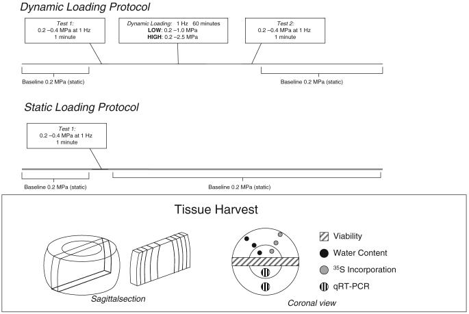
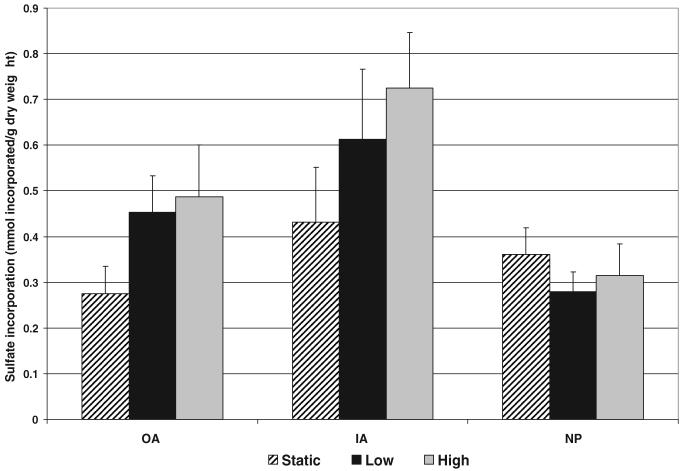
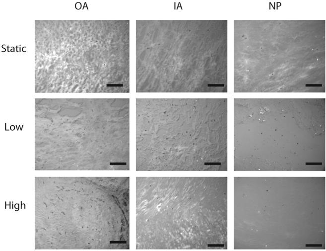
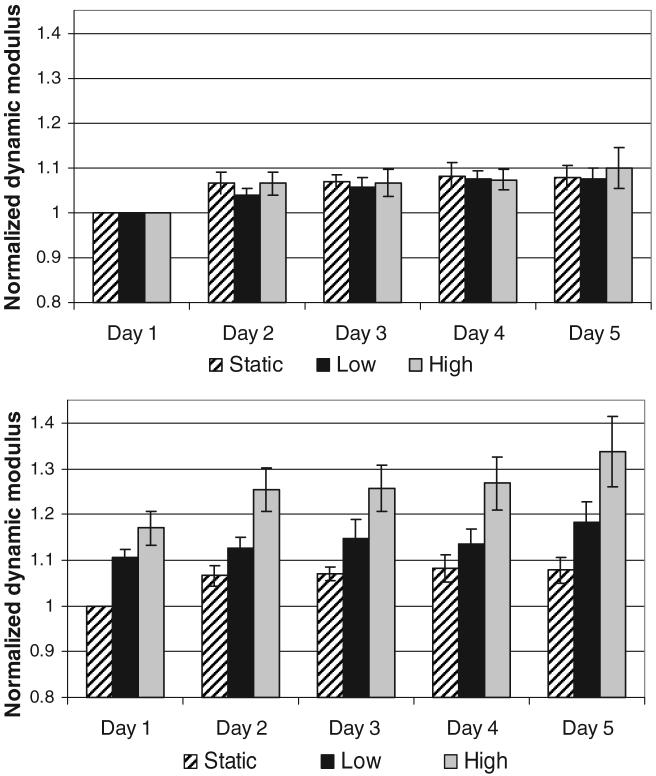
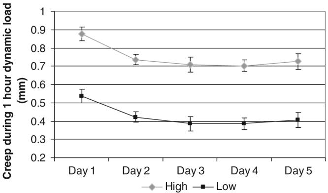
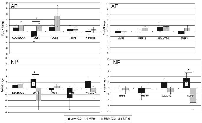
Methods: Bovine caudal intervertebral discs were placed in organ culture for 6 days and assigned to a static control or 1 of 2 dynamic compression loading protocols (0.2-1 MPa or 0.2-2.5 MPa) at 1 Hz for 1 hour for 5 days. Disc structure was assessed with measurements of dynamic modulus, creep, height loss, water content, and proteoglycan loss to the culture medium. Cellular responses were assessed through changes in cell viability, metabolism, and qRT-PCR analyses.
Results: Increasing magnitudes of compression increased disc modulus and creep; however, all mechanical parameters recovered each day. In the anulus, significant increases in gene expression for collagen I and a trend of increasing sulfate incorporation were observed. In the nucleus, increasing gene expression for collagen I and MMP3 was observed between magnitudes and between static controls and the lowest magnitude of loading.
Conclusion: Results support the hypothesis that biologic remodeling precedes damage to the intervertebral disc structure, that compression is a healthy loading condition for the disc, and further support the link between applied loading and biologic remodeling.
Figures
References
-
- Walsh AJ, Lotz JC. Biological response of the intervertebral disc to dynamic loading. J Biomech. 2004;37:329–37. - PubMed
-
- Maclean JJ, Lee CR, Alini M, et al. Anabolic and catabolic mRNA levels of the intervertebral disc vary with the magnitude and frequency of in vivo dynamic compression. J Orthop Res. 2004;22:1193–200. - PubMed
-
- Kasra M, Merryman WD, Loveless KN, et al. Frequency response of pig intervertebral disc cells subjected to dynamic hydrostatic pressure. J Orthop Res. 2006;24:1967–73. - PubMed
Publication types
MeSH terms
Substances
Grants and funding
LinkOut - more resources
Full Text Sources
Research Materials
Miscellaneous
