Review
. 2008 Jun;85(6):406-16.
doi: 10.1097/OPX.0b013e31817841e5.

Neuroprotection in glaucoma: drug-based approaches

Affiliations
Review

Neuroprotection in glaucoma: drug-based approaches

William Cheung et al. Optom Vis Sci. 2008 Jun.

Abstract

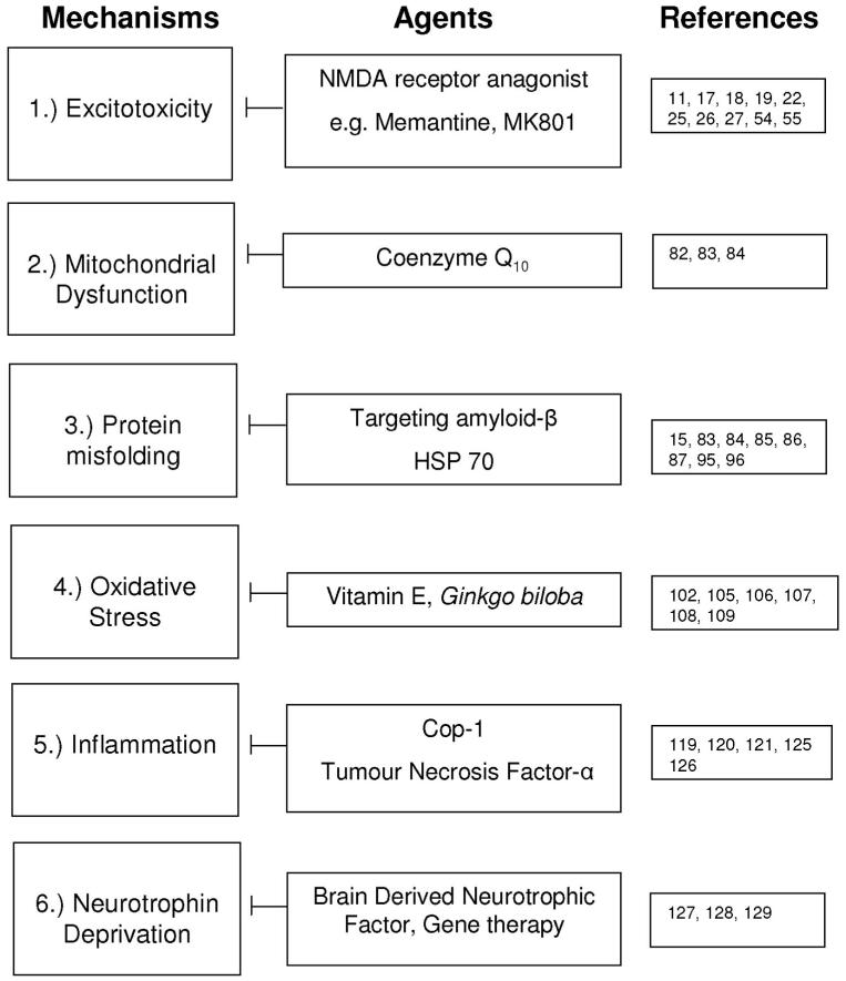
In recent years the focus of glaucoma research has shifted toward neuroprotection, as the traditional strategies of lowering intraocular pressure have been shown to be unable to prevent progressive vision loss in some glaucoma patients. As a result various neuroprotective drug-based approaches have been shown capable of reducing the death of retinal ganglion cells, which is the hallmark of glaucomatous optic neuropathy. There has been increasing evidence that glaucomatous neurodegeneration is analogous to other neurodegenerative diseases in the central nervous system, with recent work from our group elucidating a strong link between basic cellular processes in glaucoma and Alzheimer's disease. Additionally, there is a growing trend for using existing neuroprotective strategies in central nervous system diseases for the treatment of glaucoma. In fact, a trial treating patients with primary open-angle glaucoma with memantine, a drug approved for the treatment of Alzheimer's disease, has recently been completed. Results of this trial are not yet available. In this review, we will examine currently advocated neuroprotective drug-based strategies in the potential management of glaucoma.


Figures

FIGURE 1
FIGURE 1
Summary of current research strategies employed to study neuroprotection in glaucoma as previously applied in CNS disease.
FIGURE 2
FIGURE 2
A, Upon excitation by the nerve impulse, glutamate is released from the presynaptic terminal into the synaptic cleft where it binds onto the NMDA-receptors located on the postsynaptic terminal. Glutamate transporters located presynaptic terminal actively transport glutamate back into the terminal. B, Glutamate and glycine bind onto the receptors and the interaction causes transient conformational change in the channel and the depolarization of the cell resulting in the liberation of the Mg2+ which blocks the channel. The opening of the channel allows extracellular ionic molecules such as Ca2+ and Na+ to diffuse through the channel into the cell. Under normal physiological conditions, the NMDA channel is closed and is open transiently to enable the generation of a nerve impulse. C, When there is excessive glutamate present, the channel remains open causing a flood of Ca2+ and Na+ resulting in the depolarization of the mitochondrial membrane potential. Such events trigger off the release of cytochrome c which subsequently activates the caspase pathway leading toward apoptosis. A color version of this figure is available at www.optvissci.com.
FIGURE 3
FIGURE 3
Schematic diagram illustrating the role of CoQ10 in energy production in the mitochondria and its neuroprotective effects. CoQ10 transports electrons between complexes I, II, and III, and causes the augmentation of complex I in the electron transport chain (A). It is also believed to inhibit the action of NF-κB, a transcription factor responsible for inflammation, autoimmune disease, viral infection and linked with cancer (B); and finally, it inhibits the opening of the mitochondrial permeability transition pore (C). A color version of this figure is available at www.optvissci.com
FIGURE 4
FIGURE 4
Schematic diagram illustrating the formation of Aβ aggregates which ultimately leads to neuronal death. The red boxes represent three different agents which act on three different stages of Aβ pathway to block Aβ formation, deposition, and aggregation, respectively. A color version of this figure is available at www.optvissci.com.

References

    1. Cordeiro MF, Guo L, Luong V, Harding G, Wang W, Jones HE, Moss SE, Sillito AM, Fitzke FW. Real-time imaging of single nerve cell apoptosis in retinal neurodegeneration. Proc Natl Acad Sci USA. 2004;101:13352–6. - PMC - PubMed
    1. Quigley HA. Number of people with glaucoma worldwide. Br J Ophthalmol. 1996;80:389–93. - PMC - PubMed
    1. Levin LA. Retinal ganglion cells and neuroprotection for glaucoma. Surv Ophthalmol. 2003;48(suppl 1):S21–S24. - PubMed
    1. Goldberg I. Is this neuroprotective drug good for my glaucoma patients? Some key factors in clinical decision-making. Can J Ophthalmol. 2007;42:418–20. - PubMed
    1. Wein FB, Levin LA. Current understanding of neuroprotection in glaucoma. Curr Opin Ophthalmol. 2002;13:61–7. - PubMed

Substances