. 2008 Oct;117(5):457-69.
doi: 10.1007/s00412-008-0165-5. Epub 2008 Jun 3.

Plk1 regulates mitotic Aurora A function through betaTrCP-dependent degradation of hBora

Affiliations

Plk1 regulates mitotic Aurora A function through betaTrCP-dependent degradation of hBora

Eunice H Y Chan et al. Chromosoma. 2008 Oct.

Abstract

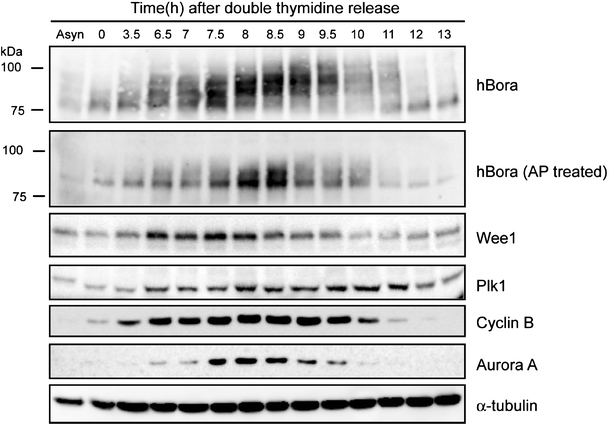
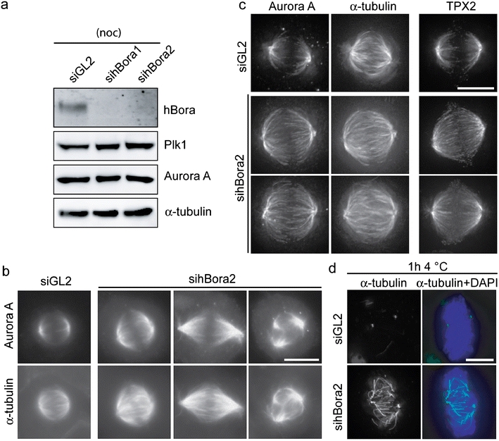
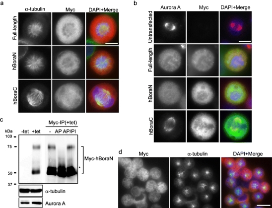
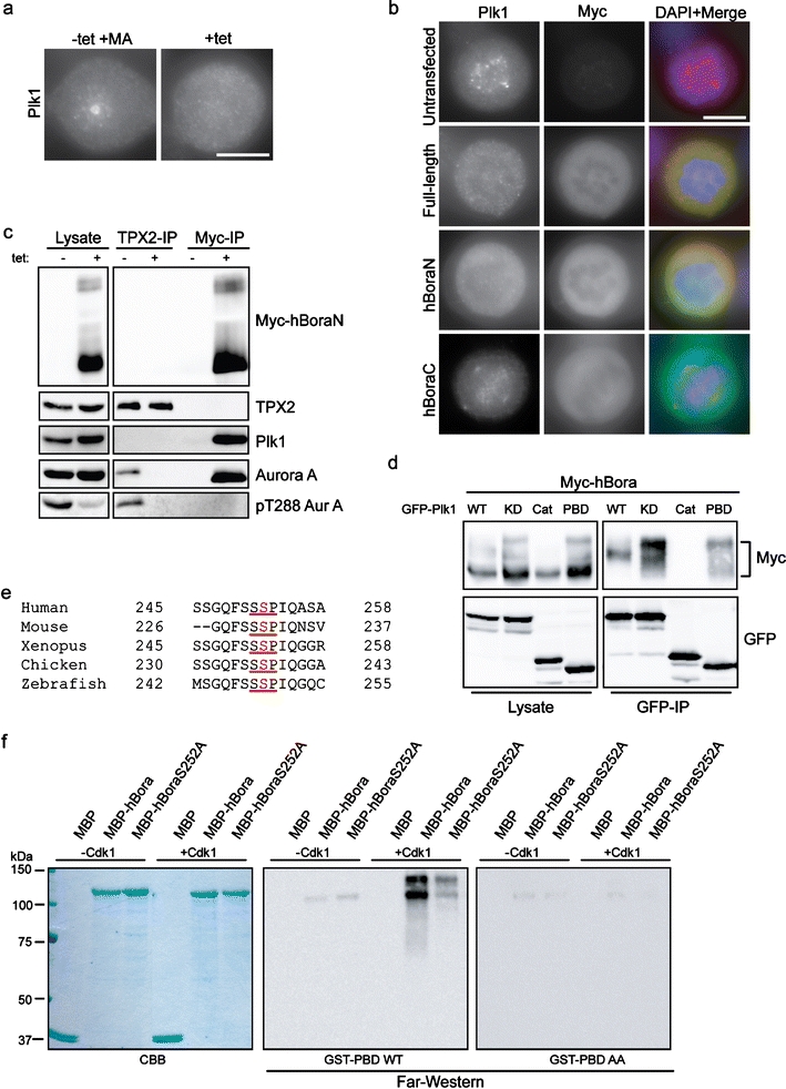
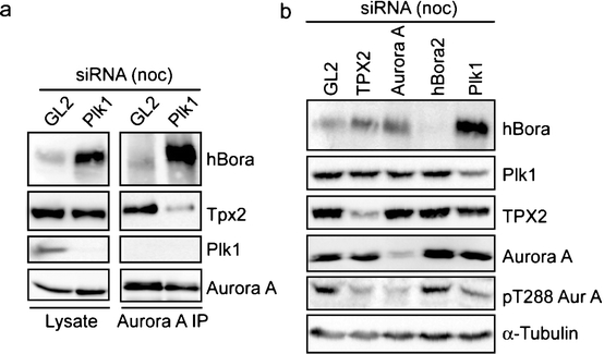
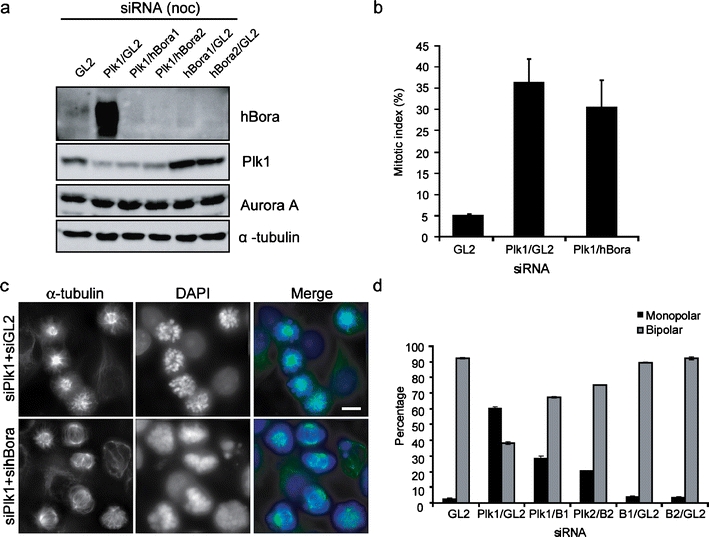
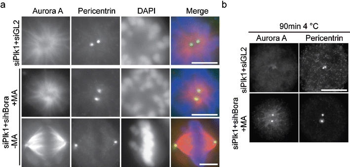
Polo-like kinase 1 (Plk1) and Aurora A play key roles in centrosome maturation, spindle assembly, and chromosome segregation during cell division. Here we show that the functions of these kinases during early mitosis are coordinated through Bora, a partner of Aurora A first identified in Drosophila. Depletion of human Bora (hBora) results in spindle defects, accompanied by increased spindle recruitment of Aurora A and its partner TPX2. Conversely, hBora overexpression induces mislocalization of Aurora A and monopolar spindle formation, reminiscent of the phenotype seen in Plk1-depleted cells. Indeed, Plk1 regulates hBora. Following Cdk1-dependent recruitment, Plk1 triggers hBora destruction by phosphorylating a recognition site for SCF(Beta-TrCP). Plk1 depletion or inhibition results in a massive accumulation of hBora, concomitant with displacement of Aurora A from spindle poles and impaired centrosome maturation, but remarkably, co-depletion of hBora partially restores Aurora A localization and bipolar spindle formation. This suggests that Plk1 controls Aurora A localization and function by regulating cellular levels of hBora.


Figures

Fig. 1
Fig. 1
hBora is cell-cycle regulated and phosphorylated during mitosis. HeLa S3 cells were synchronized by double thymidine block (G1/S phase, indicated as time 0) and released. Samples were collected at the indicated times and probed by Western blotting, using the indicated antibodies. The first lane shows a lysate from asynchronously growing cells (Asyn). Part of the lysates was treated for 30 min with alkaline phosphatase (AP) to confirm that the retarded electrophoretic mobility of hBora was phosphorylation dependent
Fig. 2
Fig. 2
Aberrant spindle formation in hBora-depleted cells. a Western blotting of mitotic HeLa S3 cells treated for 72 h with GL2 (control) siRNA oligonucleotide or two independent oligonucleotides targeting hBora (sihBora1 and 2, respectively). Membranes were probed for hBora, Plk1, Aurora A, and for α-tubulin as loading control. b HeLa S3 cells treated with GL2 (control) or hBora2 siRNA for 72 h were fixed and permeabilized with methanol. Cells were stained to detect Aurora A and α-tubulin by immunofluorescence. Bar 10 μm. c Cells (treated as in b) were processed for immunofluorescence staining with antibodies against Aurora A, α-tubulin, and TPX2. Delta Vision pictures are shown. Note increased microtubule density and increased Aurora A and TPX2 (shown in separate cells) staining on the spindles of hBora-depleted cells. Bar 10 μm. d Cells (treated as in b) were placed on ice for 1 h and then fixed and permeabilized. Cells were stained with anti-α-tubulin (green), and DNA was visualized using DAPI (blue). Bar 10 μm
Fig. 3
Fig. 3
Overexpression of hBora leads to monoastral spindle formation and Aurora A mislocalization. a, b HeLa S3 cells were transfected with the indicated Myc-tagged hBora constructs, fixed and stained with 9E10 anti-Myc (red) and α-tubulin (green; a) or with 9E10 anti-Myc (green) and Aurora A (red) antibodies (b). DNA was visualized using DAPI (blue). Bars 10 μm. c Lysates prepared from HEK293T cells expressing Myc-hBoraN upon induction with 1 μg/ml tetracyclin (+tet) for 48 h were subjected to immunoprecipitation with 9E10 anti-Myc antibody and treated with alkaline phosphatase (AP) in the presence or absence of phosphatase inhibitors (PI). Lysates and immunoprecipitated proteins were separated by sodium dodecyl sulfate polyacrylamide gel electrophoresis (SDS-PAGE) and probed by Western blotting with 9E10 anti-Myc, anti-Aurora A, and α-tubulin antibodies. Note the absence of the upper band in the sample treated with AP. The asterisk denotes the IgG heavy chain. d Cells (induced as in c) were fixed and stained with 9E10 anti-Myc (red) and α-tubulin (green) antibodies. DNA was visualized using DAPI (blue). Bar 10 μm
Fig. 4
Fig. 4
hBora interacts with Plk1 during mitosis. a Cells (induced as in Fig. 3c) were fixed and stained with an antibody against Plk1. DNA was visualized using DAPI (blue). Bar 10 μm. b HeLa S3 cells transfected with the indicated Myc-tagged hBora constructs were fixed and stained with 9E10 anti-Myc (green) and anti-Plk1 (red) antibodies. DNA was visualized using DAPI (blue). Bar 10 μm. c Lysates from HEK293T (prepared as in Fig. 3c) were subjected to immunoprecipitation with TPX2 or 9E10 anti-Myc antibodies. Lysates and immunoprecipitated proteins were separated by SDS-PAGE and probed by Western blotting with 9E10 anti-Myc, anti-TPX2 anti-Aurora A, anti-pT288 Aurora A, and α-tubulin antibodies. d HEK293T cells were co-transfected with Myc-hBora and GFP-tagged-Plk1 constructs [wild-type (WT); kinase dead (KD); catalytic domain (Cat), polo-box domain (PBD)] for 48 h, and cells were arrested with nocodazole for the last 16 h. The presence of Myc-hBora in GFP immunoprecipitates was assessed by Western blotting. e Evolutionary conservation of the putative PBD binding site in hBora (underlined). The phosphorylated serine is marked in red. Numbers refer to their position. f In vitro kinase assay was performed with Cdk1-cyclin B (or buffer for control) and the indicated proteins as substrates. The samples were subjected to SDS-PAGE, and subsequently, a far-Western ligand blotting assay using GST-PBD and GST-PBD-AA was performed. Coomassie blue staining (CBB) showed protein loading
Fig. 5
Fig. 5
Plk1 controls hBora levels. a Western blotting of mitotic HeLa S3 cells treated with Plk1 or GL2 (control) siRNA for 36 h. Lysates were subjected to immunoprecipitation with Aurora A antibody. Lysates and immunoprecipitated proteins were separated by SDS-PAGE and probed by Western blotting with anti-hBora, anti-TPX2 anti-Plk1, and anti-Aurora A antibodies. b Western blotting of mitotic HeLa S3 cells treated for 72 h with GL2 (control), TPX2, Aurora A, hBora2 siRNA oligonucleotides, or for 36 h with Plk1 siRNA oligonucleotide. Membranes were probed for hBora, Plk1, TPX2, Aurora A, pT288 Aurora A, and α-tubulin for loading control
Fig. 6
Fig. 6
hBora levels are critical for proper spindle assembly. a Western blotting of mitotic HeLa S3 cells treated for 72 h with different combinations of siRNA. Plk1 siRNA was added after 36 h of GL2 (control) or hBora depletion, and nocodazole was added for the last 12 h. Total amount of siRNA was held constant at 100 nM. Membranes were probed for hBora, Plk1, Aurora A, and for α-tubulin as loading control. b Histogram showing the mitotic indices of HeLa S3 cells (treated as in a). Results are from three individual experiments (300–350 per experiment), and bars indicate SD. c Hela S3 cells were treated with different combinations of siRNA, fixed and permeabilized, and stained with anti-α-tubulin antibody. DNA was visualized using DAPI. Bar 10 μm. Total amount of siRNA was held constant at 100 nM. d Histogram showing the percentage of mitotic cells with monopolar/ bipolar spindle in experiments performed as in c. (N = 3,150 cells per experiment), bars indicate SD
Fig. 7
Fig. 7
Plk1 regulates Aurora A localization by modulating hBora. a HeLa S3 cells, treated as in Fig. 6c and treated with or without 150 μM monastrol (MA) were fixed and stained for Aurora A (red) and pericentrin (green). DNA was visualized using DAPI. Bar 10 μm. b Same as a but cells were subjected to cold treatment for 90 min before fixation. Cells were stained with anti-Aurora A and anti-pericentrin antibodies. Note that the frequency of the observed rescue (restoration of Aurora A to spindle poles) was comparable to the frequency of restoration of bipolar spindle formation (Fig. 6d). Bar 10 μm

References

    1. Anand S, Penrhyn-Lowe S, Venkitaraman AR. AURORA-A amplification overrides the mitotic spindle assembly checkpoint, inducing resistance to Taxol. Cancer Cell. 2003;3:51–62. doi: 10.1016/S1535-6108(02)00235-0. - DOI - PubMed
    1. Barr FA, Sillje HH, Nigg EA. Polo-like kinases and the orchestration of cell division. Nat Rev Mol Cell Biol. 2004;5:429–440. doi: 10.1038/nrm1401. - DOI - PubMed
    1. Barros TP, Kinoshita K, Hyman AA, Raff JW. Aurora A activates D-TACC-Msps complexes exclusively at centrosomes to stabilize centrosomal microtubules. J Cell Biol. 2005;170:1039–1046. doi: 10.1083/jcb.200504097. - DOI - PMC - PubMed
    1. Bayliss R, Sardon T, Vernos I, Conti E. Structural basis of Aurora-A activation by TPX2 at the mitotic spindle. Mol Cell. 2003;12:851–862. doi: 10.1016/S1097-2765(03)00392-7. - DOI - PubMed
    1. Blagden SP, Glover DM. Polar expeditions—provisioning the centrosome for mitosis. Nat Cell Biol. 2003;5:505–511. doi: 10.1038/ncb0603-505. - DOI - PubMed

Publication types

MeSH terms