Review
. 2008 Aug;10(8):1343-74.
doi: 10.1089/ars.2007.1957.

Redox regulation of cell survival

Affiliations
Review

Redox regulation of cell survival

Dunyaporn Trachootham et al. Antioxid Redox Signal. 2008 Aug.

Abstract

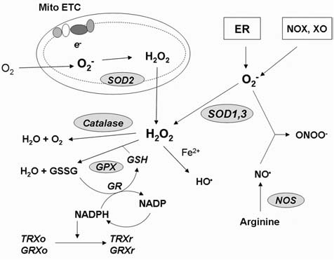
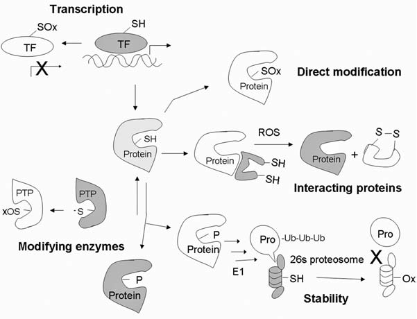
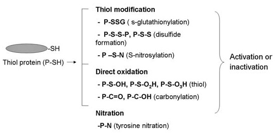
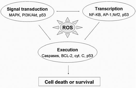
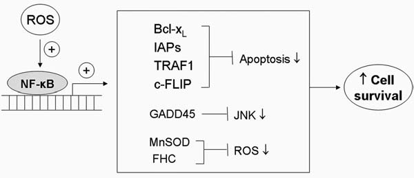
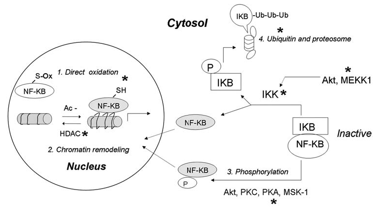
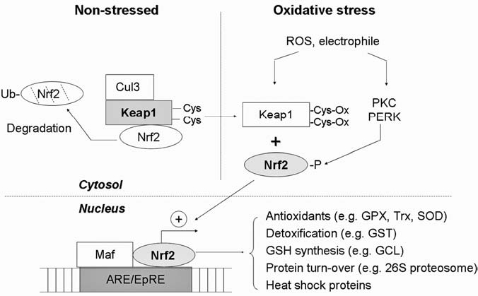
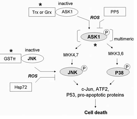
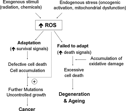
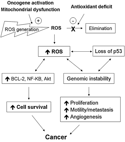
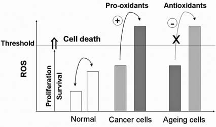
Reactive oxygen species (ROS) and reactive nitrogen species (RNS) play important roles in regulation of cell survival. In general, moderate levels of ROS/RNS may function as signals to promote cell proliferation and survival, whereas severe increase of ROS/RNS can induce cell death. Under physiologic conditions, the balance between generation and elimination of ROS/RNS maintains the proper function of redox-sensitive signaling proteins. Normally, the redox homeostasis ensures that the cells respond properly to endogenous and exogenous stimuli. However, when the redox homeostasis is disturbed, oxidative stress may lead to aberrant cell death and contribute to disease development. This review focuses on the roles of key transcription factors, signal-transduction pathways, and cell-death regulators in affecting cell survival, and how the redox systems regulate the functions of these molecules. The current understanding of how disturbance in redox homeostasis may affect cell death and contribute to the development of diseases such as cancer and degenerative disorders is reviewed. We also discuss how the basic knowledge on redox regulation of cell survival can be used to develop strategies for the treatment or prevention of those diseases.


Figures

FIG. 1.
FIG. 1.
Redox homeostasis. Major sites of cellular ROS generation include the mitochondrial electron transport chain (Mito ETC), the endoplasmic reticulum (ER) system, and the NAD(P)H oxidase (NOX) complex. Nitric oxide synthases (NOS) are key enzymes for production of NO. Major ROS-scavenging enzymes are highlighted in grey. GSH and NAPDH play roles in maintaining the reduced cellular redox state. GPX, glutathione peroxidase; GR, glutathione reductase; TRXo, thioredoxin (oxidized); TRXr, thioredoxin (reduced); GRXo, glutaredoxin (oxidized); GRXr, glutaredoxin (reduced); HO·, hydroxyl radical; NO·, nitric oxide; ONOO, peroxynitrite; SOD, superoxide dismutase; GSH, reduced glutathione; GSSG, oxidized glutathione; NADPH, reduced nicotinamide adenine dinucleotide phosphate; XO, xanthine oxidase.
FIG. 2.
FIG. 2.
Redox-mediated mechanisms that regulate protein functions. Protein expression can be regulated through redox modification of transcription factors. Oxidation of Cys at or near the DNA-binding site may disrupt the transactivation activity. Newly synthesized protein can be directly modified by oxidation of amino acids such as Cys, Tyr, and Met, resulting in alteration of the protein functions. Certain proteins are stabilized by their redox-sensitive interacting proteins. Modification of the interacting proteins can dissociate the complex and allow activation of the functional proteins. Posttranslational modifications such as phosphorylation can either activate or inhibit protein functions. Phosphatases, which are responsible for dephosphorylation, can be oxidatively inactivated, promoting phosphorylation of proteins. Stability of signaling proteins determines both the level and duration of their functional effects. Most proteins can be degraded through the ubiquitin–proteosome system. Ubiquitin-activating enzyme E1 and proteosome 26S and 20S can be inactivated under oxidative stress. TF, transcription factor; -SH, reduced thiol; SOx, oxidized thiol; PTP, protein tyrosine phosphatase; ub, ubiquitin. X, inhibition; white, inactive state; light grey, partially activated; dark grey, fully activated molecules.
FIG. 3.
FIG. 3.
Oxidative modification of proteins. Protein can be oxidatively modified by multiple types of modifications, and the consequence can be either activation or inactivation of protein functions. Cys in thiol proteins is a major target, which could be modified by reversible S-glutathionylation, disulfide formation, S-nitrosylation, or formation of sulfinic, sulfenic, and sulfonic acid derivatives. Nitration of Tyr is known to modulate the function of multiple kinases. P, protein.
FIG. 4.
FIG. 4.
Redox-sensitive signaling pathways for regulation of cell survival. The redox system can regulate the cell-fate decision through regulations of many functional proteins involving cell life-or-death decisions. Many of those signaling proteins are redox sensitive, which controls survival at the levels of signal transduction, transcriptional regulation, or execution. Examples of the key redox-sensitive molecules involved at each level are indicated. The possible crosstalk among these regulators/executors is indicated by arrows. Signal transduction may involve in transcriptional regulation, and p53 is a redox-sensitive molecule that affects cell survival at all three levels. Therefore, oxidative stress not only serves as a type of stimulus to trigger stress-response signal-transduction pathways, but also can modulate cell death/survival through direct oxidative modification of those signal molecules.
FIG. 5.
FIG. 5.
Role of NF-κB in cell survival. NF-κB functions as a transcription factor regulating the expression of multiple genes. Activation of NF-κB by stimuli such as oxidative stress or cytokines promotes increased expression of antiapoptotic proteins such as Bcl-xL and XIAP, which suppress the execution phase of cell death. Induction of GADD45 leads to inhibition of JNK and prevents JNK-induced apoptosis. NF-κB also promotes the expression of antioxidant genes such as MnSOD, which plays a major role in scavenging mitochondria superoxide and in maintaining redox homeostasis. Overall, the activation of NF-κB by ROS leads to inhibition of apoptosis, redox rebalance, and enhanced cell survival.
FIG. 6.
FIG. 6.
Redox regulation of NF-κB. The function of NF-κB can be activated or inhibited through various redox-mediated mechanisms at multiple levels of the activation pathways. In the nucleus, direct oxidation of Cys in the DNA-binding domain can inhibit NF-κB-DNA-binding activity. In contrast, enzyme histone deacetylase (HDAC), which catalyzes the removal of an acetyl (Ac-) group from histone, can be inactivated by oxidative stress, allowing histone acetylation, chromatin uncoiling, and increased accessibility for NF-κB. In cytosol, activation of NF-κB can be regulated through phosphorylation of NF-κB itself or phosphorylation of its inhibitor IκB. Normally, NF-κB and IκB form a complex, which is sequestered in cytosol. Increased ROS can activate IκB-kinase (IκK) either directly through redox modification of IκK, or indirectly through activation of Akt and/or MEKK1, which then phosphorylates and activates IκK. Active IκK phosphorylates IκB and liberates active NF-κB from the complex to translocate to the nucleus. Phosphorylated IκB undergoes ubiquitination and degradation by proteosomes. Because the proteosome system is also redox sensitive, ROS can also regulate NF-κB activity by affecting the stability of IκB. Furthermore, phosphorylation of NF-κB by certain kinases may dissociate NF-κB from IκB and promote its nuclear translocation. Grey, Active forms of the proteins. *Major target molecules of redox regulation.
FIG. 7.
FIG. 7.
Redox regulation of Nrf2. In unstressed cells, Nrf2 is sequestered in cytosol by Keap1, which functions as an adaptor for Cul3 (a ubiquitin E3 ligase) to target Nrf2 for ubiquitination and degradation. On oxidative stress or electrophilic stimuli, Nrf2 is activated via two mechanisms: (a) thiol oxidation of Keap1 and (b) phosphorylation of Nrf2 by kinases such as PKC or PERK. These cause release of Nrf2 from the inactive complex. The free Nrf2 is translocated to the nucleus, where it forms a heterodimer with Maf proteins and then binds to the antioxidant-responsive element or electrophile-responsive element (ARE/EpRE). The active binding triggers transcription of multiple target genes that encode antioxidants, glutathione synthesis enzymes, proteosomes, and heat-shock proteins. Grey, Active forms of the molecules. GPX, glutathione peroxidase; Trx, thioredoxin; SOD, superoxide dismutase; GCL, glutamylcysteine ligase, GST; glutathione-S-transferase.
FIG. 8.
FIG. 8.
Redox regulation of stress-responsive kinase (SAPK) signaling pathways. In most cases, activation of the SAPK pathway transduces an oxidative stress signal to cell death. Under nonstressed conditions, apoptosis-regulating signal kinase 1(ASK1) is inhibited by the reduced form of thioredoxin (Trx) or glutaredoxin (Grx). Increased oxidative stress causes oxidation of Trx and Grx and releases ASK1 to form an active multimeric complex with proper trans- or autophosphorylation. The activation of ASK1 subsequently leads to activation of c-Jun N-terminal kinase (JNK) and p38-MAPK, resulting in induction of cell death. JNK can also be inhibited by complex formation with glutathione S-transferase-π (GST-π) under nonstressed conditions, and can be activated by ROS in a similar fashion as ASK1. Negative regulatory molecules include the Ser/Thr phosphatase 5 (PP5), which inhibits ASK1 kinase activity by causing its dephosphorylation, and heat-shock protein 72 (Hsp72), which inhibits JNK activity. *Site of redox regulation. Grey, Active forms of the proteins.
FIG. 9.
FIG. 9.
Redox regulation of the PI3K/Akt signaling pathway. PI3K/Akt transduces the signal for cell survival mainly through phosphorylation of target molecules by Akt. This results in inactivation of proapoptotic proteins and activation of transcription factors, which target antiapoptotic proteins. Under oxidative stress, this pathway is activated by oxidative inactivation of phosphatases [i.e., protein tyrosine phosphatases (PTPases) and PTEN], allowing constitutive activation of tyrosine kinase receptor and PI3K. However, direct oxidative modification of PI3K and Akt can result in their inactivation and compromise the survival signals. Furthermore, the PI3K/Akt pathway can also promote cellular production of ROS through activation of Rac and NADPH oxidase (NOX). *Site of redox regulation. Grey, Active forms of the proteins; TORC2, mTOR complex 2.
FIG. 10.
FIG. 10.
Redox regulation at the execution level. Apoptosis can be triggered through extrinsic or intrinsic pathways. External stimuli such as TNF-α or Fas ligand binds to death receptor and transduces the signal into activation of caspase-8, leading to initiation of the extrinsic pathway. Intrinsic signals, such as DNA damage and oxidative stress, can transduce the death signal by causing release of cytochrome c from mitochondria to cytosol, followed by activation of caspase-9 through formation of apoptosome (Apaf-1, cytochrome c, pro-caspase 9, and dATP). Active caspase 8 and caspase 9 can further cleave procaspase-3, producing an active fragment of caspase-3, which cleaves its protein substrates such as PARP, resulting in apoptosis. To keep apoptosis in check, Bcl-2 family proteins play an important role in regulation of mitochondria membrane permeability and cytochrome c release. Antiapoptotic proteins such as Bcl-2 prevent apoptosis through both direct interaction with proapoptotic proteins such as Bax and indirect control of oxidative stress via maintenance of a reducing environment. Grey*, Potential sites of redox regulation. Car, cardiolipin; Cyt c, cytochrome c; casp 3, caspase 3.
FIG. 11.
FIG. 11.
Integration of redox signaling. An increase of reactive oxygen species (ROS) can result in either cell survival or cell death, depending on the integration of the proapoptotic and antiapoptotic signals. The levels and durations of ROS stress determine the activation or inhibition of each signal-transduction pathway. The crosstalk between PI3K/Akt and stress-responsive MAPK pathway (SAPK) serves as an example. A low level or transient increase of ROS can activate PI3K/Akt, leading to cell survival through NF-κB. The predominant survival signals of the Akt and NF-κB pathways prevent cell death by inhibiting ASK1 and JNK, respectively. However, a high level of ROS causes a sustained activation of the ASK1-JNK cascade and inactivation of PI3K/Akt and NF-κB due to protein oxidation, leading to cell death.
FIG. 12.
FIG. 12.
Regulation of cell fate by p53. Different levels of ROS stress activate p53, leading to various outcomes. Activation of p53 by a low level of ROS stress promotes cell survival, whereas severe ROS stress activates p53 and causes cell death. It is possible that the activated p53 might interact with different factors (indicated as “X” and “Y”) under different levels of ROS stress, leading to activation of distinct sets of target genes (see text for details).
FIG. 13.
FIG. 13.
Disturbance of redox homeostasis and pathogenesis of diseases. Alterations in redox homeostasis by exogenous stimuli or endogenous stress or both can result in increased oxidative stress with elevated cellular ROS. The ability to adapt to such ROS stress determines the overall fates of the cells. A successful adaptation to increased survival signals in combination with further ROS-mediated mutations and loss of critical regulatory mechanisms lead to defective cell death and aberrant proliferation. These may contribute to development of cancer. A failure in adaptation to the ROS stress while the cells accumulate oxidative DNA, protein, and lipid damage product may result in excessive cell death, leading to degenerative disorders and aging.
FIG. 14.
FIG. 14.
Redox alterations and cancer development. Disturbance of redox homeostasis by an increase in ROS production (due to oncogenic stimulation, mitochondrial dysfunction, etc.) or a decrease in ROS elimination (due to a deficit or dysfunction of the antioxidant system) can lead to elevated ROS. The increased ROS stress can induce DNA mutations and genetic instability, including a loss of tumor-suppressor genes such as p53. The loss of p53 function can in turn further contribute to mitochondrial dysfunction, ROS generation, and genomic instability, forming a vicious cycle. ROS stress may also induce the expression of prosurvival factors and certain ROS-scavenging proteins, which would enable the cells to adapt and survive under oxidative stress. The ROS-induced mutations and genetic instability further enhance the chance for selection of cells with malignant phenotypes (an increase in proliferation, survival capacity, cell motility, and angiogenesis), leading to development of cancer.
FIG. 15.
FIG. 15.
Redox homeostasis and strategies to modulate redox dynamics for potential therapeutic applications. Under physiologic conditions, normal cells maintain redox homeostasis by controlling the proper balance between ROS generation and elimination. The redox dynamics may fluctuate within a tolerable range. An increase of ROS may promote cell proliferation and survival, as in the case of many cancer cells. However, when the increase of ROS reaches a critical level (the threshold), it may overwhelm the cellular antioxidant capacity and trigger the cell-death process. Chronic ROS stress may cause accumulation of damage to a level that induces cell death. This is thought to be a mechanism contributing to neural degenerative diseases and aging. For therapeutic purposes, it is possible to use agents that promote ROS generation or inhibit the cellular antioxidant system to trigger cancer cell death by pushing the ROS above the threshold level. In contrast, antioxidants may be used to prevent cells from oxidative damage and delay aging and the neurodegenerative process.

References

    1. Adler V. Yin Z. Fuchs SY. Benezra M. Rosario L. Tew KD. Pincus MR. Sardana M. Henderson CJ. Wolf CR. Davis RJ. Ronai Z. Regulation of JNK signaling by GSTp. EMBO J. 1999;18:1321–1334. - PMC - PubMed
    1. Alexandre J. Nicco C. Chereau C. Laurent A. Weill B. Goldwasser F. Batteux F. Improvement of the therapeutic index of anticancer drugs by the superoxide dismutase mimic mangafodipir. J Natl Cancer Inst. 2006;98:236–244. - PubMed
    1. Angel P. Karin M. The role of Jun, Fos and the AP-1 complex in cell-proliferation and transformation. Biochim Biophys Acta. 1991;1072:129–157. - PubMed
    1. O'Brian CA. Chu F. Review: post-translational disulfide modifications in cell signaling: role of inter-protein, intra-protein, S-glutathionyl, and S-cysteaminyl disulfide modifications in signal transmission. Free Radic Res. 2005;39:471–480. - PubMed
    1. Arakawa M. Ito Y. N-acetylcysteine and neurodegenerative diseases: basic and clinical pharmacology. Cerebellum. 2007;6:308–314. - PMC - PubMed

Publication types

Substances

LinkOut - more resources