. 2008;10(3):R50.
doi: 10.1186/bcr2104. Epub 2008 Jun 3.

Mammary carcinoma behavior is programmed in the precancer stem cell

Affiliations

Mammary carcinoma behavior is programmed in the precancer stem cell

Patrizia Damonte et al. Breast Cancer Res. 2008.

Abstract

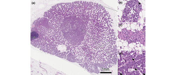
Introduction: The 'MINO' (mammary intraepithelial neoplasia outgrowth) mouse model of ductal carcinoma in situ (DCIS) consists of six lines with distinct morphologic phenotypes and behavior, each meeting experimentally defined criteria for 'precancer'. Specifically, these lines grow orthotopically in cleared mammary fat pads and consistently progress to an invasive phenotype that is capable of ectopic growth. Transition to carcinoma has a consistent latency for each line, and three of the lines also exhibit pulmonary metastatic potential.

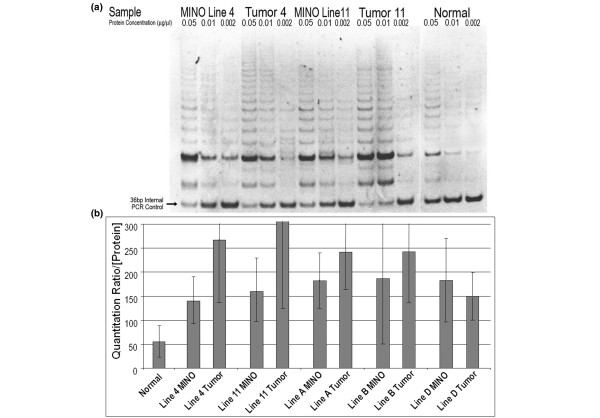
Methods: Gland cleared orthotopic transplanted precancer MINO tissues were analyzed by bacterial artifical chromosome and oligo array comparative genomic hybridization, microsatellite PCR, and telomerase repeat amplification assay. MINO cells were dissociated and cultured in three dimensional culture and transplanted in syngeneic gland cleared mammary fat pads.

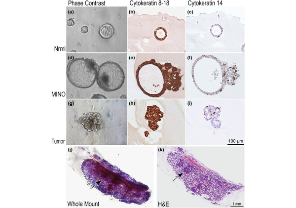
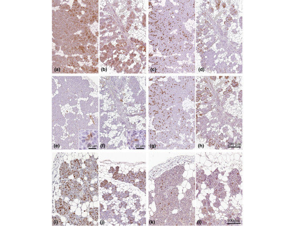
Results: Comparative genomic hybridization shows that the precancer and invasive tumors are genetically stable, with low level changes including whole chromosome gains in some lines. No changes are associated with progression, although spontaneous focal amplifications and deletions were detected occasionally. Microsatellite analysis shows a low frequency of alterations that are predominantly permanent within a MINO line. Telomerase activity is increased in both the MINO and the derived tumors when compared with normal mouse mammary gland. Dissociation of the precancer lesion cells and three dimensional 'spheroid' culture of single cells reveals a bipotential for myoepithelial and luminal differentiation and the formation of unique three-dimensional 'MINOspheres'. These MINOspheres exhibit features that are intermediate between spheroids that are derived from normal and carcinoma cells. Transplantation of a single cell derived MINOsphere recapitulates the outgrowth of the precancer morphology and progression to carcinoma.

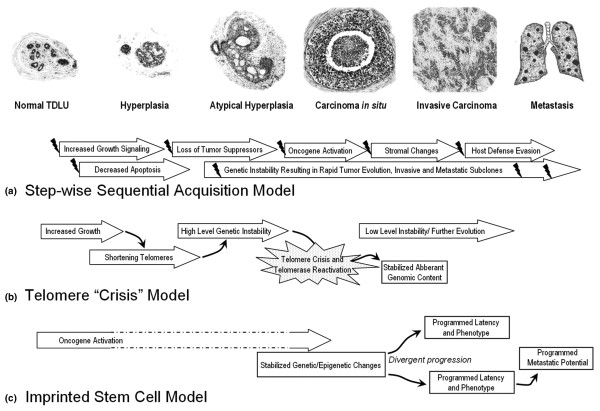
Conclusion: These data establish a precancer 'stem' cell that is capable of self-renewal and multilineage differentiation as the origin of invasive cancer. Within the context of this model, these cells have programmed potential for latency and metastasis that does not appear to require sequential genetic 'hits' for transformation.


Figures

Figure 1
Figure 1
Histomorphology of the MINO model mammary precancer with early invasive carcinoma. (a) Low-power hematoxylin and eosin histomorphology of the MINO precancer filling the precleared fat pad. Intact gland is seen for comparison in the bottom left corner. The lymph node is seen (LN) at the left, and no invasion of the node by precancer tissue is seen. (b) The edge of the precancer is seen at higher power and is similar to a normal developing mammary gland terminal end bud with mitoses (arrowhead) and remodeling via apoptosis to clear the luminal space. In the area of precancer in the center of the growth there is an organized relationship between the transplanted MINO tissue and the host stroma. (c) The MINO cells differentiate to form acinar and ductal structures with high intralesional cell heterogeneity. (d) In this heterogeneous differentiated zone of precancer tissue, an area of transformation to invasive carcinoma is characterized by increased mitoses (arrowheads) and much less residual host stromal tissue. MINO, mammary intraepithelial neoplasia outgrowth.
Figure 2
Figure 2
Telomerase activity of MINO and tumor tissues. Telomerase activity by telomerase repeat amplification protocol (TRAP) assay. Representative ethidium stained polyacrylamide gel electrophoresis of the PCR products resulting from protein extract incubation with artificial telomere repeat template showing a bright 50 bp band with 6 bp increasing length ladder. At the bottom of the gel is a 36 bp internal PCR control, with PCR optimized to be semi-competitive with the extended telomere repeats produced by the telomerase protein derived from each sample. The protein samples are diluted in buffer and then added to the reaction mixture to provide a final reaction mixture concentration as listed (top of each lane). Matched MINO and tumor samples were tested. (a) Lines 4w4 and 4w11 are shown, with normal mouse mammary epithelium control (separated from the stroma by partial tissue dissociation and centrifugation) shown in the right three lanes. (b) Quantitation by comparing the band intensity with the internal control for multiple samples is shown, with standard deviation of the mean depicted for each bar. Line 11 tumor had the highest levels, at a mean of 500 (not shown), and all samples except for line B MINO were statistically significantly different from the normal control. There was a high level of variability between assay runs, depicted by the size of the error bars, but the qualitative data were clear, as shown in the gel (panel a). bp, base pairs; MINO, mammary intraepithelial neoplasia outgrowth; TRAP, telomerase repeat amplification protocol.
Figure 3
Figure 3
Three-dimensional culture of single cells from normal prelactating mammary gland, MINO precancer, and invasive carcinoma. (a, d, g) Inverted microscope phase contrast images as well as histologic images generated from paraffin-embedded 4 μm sections of the three-dimensional cultures stained by immunohistochemistry with (b, e, h) CK8–18 or (c, f, i) CK14 are shown. The magnification scale (lower right panel i) is identical for all histology panels and approximate for the inverted microscopy photographs. CK8–18 confirms that the major cell population is a luminal phenotype, but CK14 shows that there is also myoepithelial differentiation of single cells, documenting bipotential of the individual cells giving rise to these three-dimensional structures. A single MINOsphere from line 4w4 was transplanted from the three dimensional culture into the gland cleared fat pad of a 3-week-old female FVB/n mouse. The mammary gland was removed 10 weeks after transplant and a whole mount mammary gland preparation was made with hematoxylin stain to visualize cell density (j). After photography, the same gland was processed for histologic sectioning, and the resulting 4 μm hematoxylin and eosin stained tissue section is shown (k). At least three foci of tumor are seen in the differentiation zone of the MINO. One is indicated by the arrow. CK, cytokeratin; MINO, mammary intraepithelial neoplasia outgrowth.
Figure 4
Figure 4
Immunohistochemistry profile of MINOsphere-derived outgrowth versus the original MINO tissue outgrowth. The outgrowth derived from a MINOsphere transplant (a, c, e, g, i, k) shows analogous expression compared with the MINO tissue outgrowth from the donor (b, d, f, h, j, m) for the following: luminal epithelial, CK8–18 (panels a and b); basal epithelial/myoepithelial, CK14 (panels c and d) and CK5 (panels g and h); myoepithelial/smooth muscle, SMA (panels e and f insets show higher magnification of SMA-positive cells within the epithelial layers); and proliferation/apoptosis, Ki67 (panels I and j)/caspase3 (panels k and l). Panels a to h are of identical magnification, with the 200 μm scale bar shown (panel h). Panels i to l are identical magnification with the 100 μm scale bar shown (panel l). CK, cytokeratin; MINO, mammary intraepithelial neoplasia outgrowth; SMA, smooth muscle actin.
Figure 5
Figure 5
Conceptual models of breast cancer progression. (a) Sequential acquisition of molecular alterations with selection of advantageous 'hits' and corresponding morphologic progression (modified from Burstein and coworkers [4]). Individual 'hits' are depicted with small 'lightening bolts'. (b) Hyperplasia results in shortening telomeres, rapidly increasing genetic instability during 'telomere crisis', and relative stability after telomerase reactivation (modified from Chin and coworkers [15]). Individual cells reactivating telomerase and with a 'fit' genetic profile give rise to the carcinoma in situ (DCIS) and then the invasive carcinoma. (c) Genetically stable precancer stem cells are initiated via oncogene activation with divergent behavior programmed via epigenetic encoding and possible but not required genetic content changes. Intermediate morphologic and molecular events are not required for progression. These cells give rise to the DCIS and have an innate latency to invasive carcinoma and an innate metastatic potential.

References

    1. Page DL, Dupont WD. Premalignant conditions and markers of elevated risk in the breast and their management. Surg Clin North Am. 1990;70:831–851. - PubMed
    1. Dupont WD, Page DL. Risk factors for breast cancer in women with proliferative breast disease. N Engl J Med. 1985;312:146–151. - PubMed
    1. Page DL, Dupont WD, Rogers LW, Landenberger M. Intraductal carcinoma of the breast: follow-up after biopsy only. Cancer. 1982;49:751–758. doi: 10.1002/1097-0142(19820215)49:4<751::AID-CNCR2820490426>3.0.CO;2-Y. - DOI - PubMed
    1. Burstein HJ, Polyak K, Wong JS, Lester SC, Kaelin CM. Ductal carcinoma in situ of the breast. N Engl J Med. 2004;350:1430–1441. doi: 10.1056/NEJMra031301. - DOI - PubMed
    1. Boland GP, Chan KC, Knox WF, Roberts SA, Bundred NJ. Value of the Van Nuys Prognostic Index in prediction of recurrence of ductal carcinoma in situ after breast-conserving surgery. Br J Surg. 2003;90:426–432. doi: 10.1002/bjs.4051. - DOI - PubMed

Publication types