. 2008 Aug;135(2):529-38.
doi: 10.1053/j.gastro.2008.04.020. Epub 2008 Apr 22.

Toll-like receptor signaling in small intestinal epithelium promotes B-cell recruitment and IgA production in lamina propria

Affiliations

Toll-like receptor signaling in small intestinal epithelium promotes B-cell recruitment and IgA production in lamina propria

Limin Shang et al. Gastroenterology. 2008 Aug.

Abstract

Background & aims: Several lines of evidence support a role for Toll-like receptor (TLR) signaling to protect the intestine from pathogenic infection. We hypothesized that TLR signaling at the level of the intestinal epithelium is critical for mucosal immune responses.

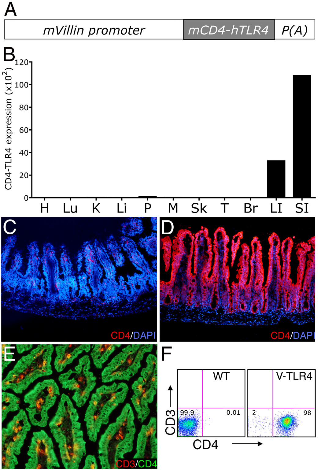
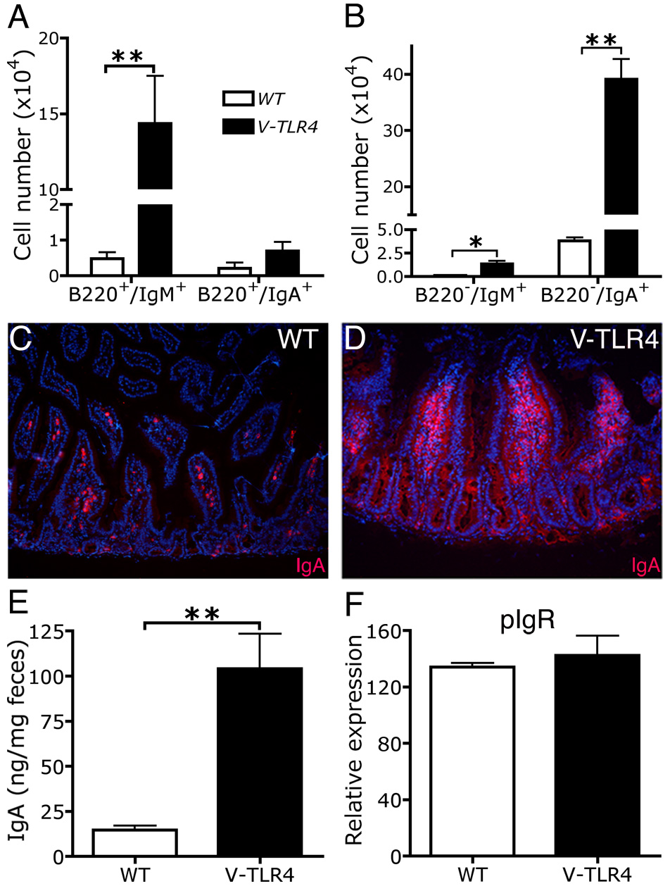
Methods: We generated transgenic mice that express a constitutively active form of TLR4 in the intestinal epithelium (V-TLR4 mice). Lamina propria cellularity was evaluated by immunostaining and flow cytometry. Immunoglobulin (Ig) A levels in the stool and serum were measured by enzyme-linked immunosorbent assay. Chemokine and cytokine expression were analyzed by quantitative polymerase chain reaction and enzyme-linked immunosorbent assay.

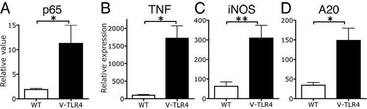
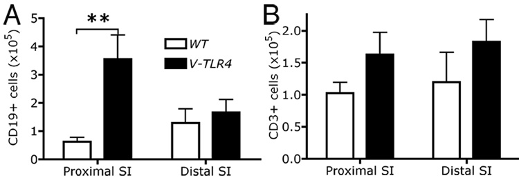
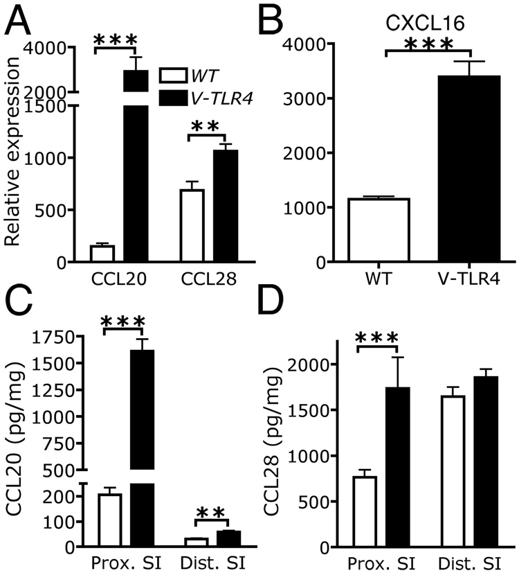
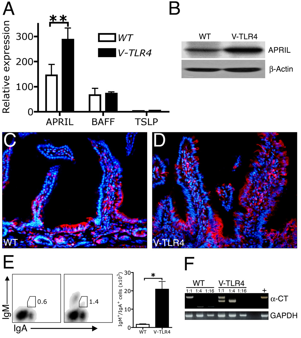
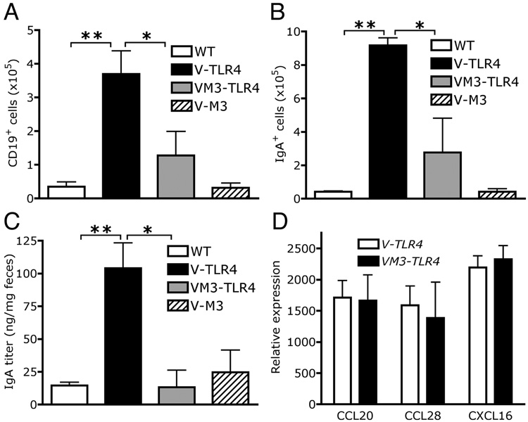
Results: V-TLR4 transgenic mice reproduced normally and had a normal life span. Constitutive activity of TLR4 in the intestinal epithelium promoted recruitment of B cells and an increase in fecal IgA levels. Intestinal epithelial cells of V-TLR4 mice expressed higher levels of CCL20 and CCL28, chemokines known to be involved in B-cell recruitment, and of a proliferation-inducing ligand (APRIL), a cytokine that promotes T-cell-independent class switching of B cells to IgA. The changes in B-cell numbers and IgA levels were blocked by simultaneous expression in intestinal epithelial cells of M3, a herpes virus protein that binds and inhibits multiple chemokines.

Conclusions: TLR signaling in the intestinal epithelial cells significantly elevated the production of IgA in the intestine. This effect was mediated by TLR-induced expression of a specific set of chemokines and cytokines that promoted both recruitment of B cells into the lamina propria and IgA class switching of B cells.


Conflict of interest statement

Statement: There is no conflict of interest to disclose

Figures

Figure 1
Figure 1. Expression of the CD4-TLR4 transgene in V-TLR4 mice
A) Diagram of the V-TLR4 transgene. The transgene encodes a fusion protein containing the mouse CD4 (mCD4) extracellular domain and human TLR4 signaling domain and is driven by the mouse villin promoter (mVillin). p(A) represents SV40 poly A sequences. B) CD4-TLR4 mRNA expression in different tissues of V-TLR4 mice. The values were standardized to ubiquitin levels in each sample. H: Heart; Lu: Lung; K: kidney; Li: liver; P: Pancreas; M: Skeletal Muscle; Sk: Skin; T: Testis; Br: Brain; LI: Large intestine; SI: Small intestine. C–D). Representative immunostaining for CD4 (red) and DAPI (blue) in small intestine of WT (C) and V-TLR4 (D) mice. E) Representative immunostaining for CD3 (red) and CD4 (green) in small intestine of V-TLR4 mice. Note that the transgene expression is restricted to epithelial cells. F) FACS analysis of CD4-TLR4 expression in epithelial cells isolated from small intestine. Cells were gated on the CD45/PI.
Figure 2
Figure 2. Activation of the TLR signaling pathway in IECs of V-TLR4 mice
A) DNA binding ELISA analysis of p65 in IECs isolated from the small intestine of WT and V-TLR4 mice (n=10/group, *p<0.05). B–D). Relative mRNA expression of TNFα (B), iNOS (C), and A20 (D) in IECs isolated from small intestine of WT and V-TLR4 mice (n=3/group; * p<0.05, ** p<0.01).
Figure 3
Figure 3. Transgene expression in IECs increases the number of B cells in the lamina propria
A–B) FACS analysis of CD19+ B cells (A) and CD3+ T cells (B) in lamina propria of WT and V-TLR4 mice. Values indicate the absolute number of each subset (n=4/group). Note the increased number of B cells in the proximal small bowel of V-TLR4 mice (** p<0.01).
Figure 4
Figure 4. Increased expression of chemokines in the proximal small intestine of V-TLR4 mice
A–B) Q-PCR analysis of CCL20, CCL28 (A) and CXCL16 (B) expression in the proximal small intestine of WT and V-TLR4 mice (n=7/group; ** p<0.01, *** p<0.001). C–D) CCL20 (C) and CCL28 (D) protein levels in small intestine of V-TLR4 mice and WT mice (WT n=12, V-TLR4 n=15; *** p< 0.001).
Figure 5
Figure 5. Increased IgA production in the lamina propria of V-TLR4 mice
A–B). FACS analysis of B cell (A) and plasma cell subsets (B) in lamina propria of proximal small intestine of WT and V-TLR4 mice (WT n=3, V-TLR4 n=4; * p< 0.05, ** p< 0.01). C & D). Immunostaining of IgA+ cells in duodenal lamina propria of WT (C) and V-TLR4 (D) mice. E). IgA levels in feces of WT (n=24) and V-TLR4 (n=50) mice (** p<0.01). F). Q-PCR analysis of pIgR expression in IECs of WT and V-TLR4 mice (n=4/group).
Figure 6
Figure 6. Increase in APRIL expression in IECs and B cell class switching to IgA in V-TLR4 mice
A). Q-PCR analysis of APRIL, BAFF and TSLP expression in proximal small intestinal epithelial cells of WT and V-TLR4 mice (n=3/group; **, p<0.01). B). Western blot of APRIL protein in proximal small intestinal epithelial cells of WT and V-TLR4 mice. C & D). Immunostaining of APRIL in duodenum of WT (C) and V-TLR4 (D) mice (red, APRIL; blue, DAPI). E). FACS analysis of IgM+/IgA+ cells in lamina propria of proximal small intestine of WT and V-TLR4 mice (WT n=3, V-TLR4 n=4; * p< 0.05). F). RT-PCR analysis of post switch circular transcript (α-CT) expression in lamina propria B cells from WT and V-TLR4 mice. Data represented pooled B cells from 20 WT or 10 V-TLR4 mice. Different bands represent different splice variants of α-CT as previously described .
Figure 7
Figure 7. Blockade of B cell recruitment and IgA production in the lamina propria of V-TLR4 mice by co-expression of chemokine binding protein M3
A). Absolute number of CD19+ B cells in the lamina propria of WT, V-TLR4, V-M3 and double transgenic mice (n=4/group; ** p<0.01, * p<0.05). B). Absolute number of IgA+ cells in the lamina propria of the proximal small intestine of WT, V-TLR4, V-M3 and double transgenic mice (n=4/group; ** p<0.01, * p<0.05). C). IgA levels in feces of WT, V-TLR4, V-M3 and double transgenic mice (WT n=36, V-TLR4 n=51, V-M3 n=18, V-M3/V-TLR4 double TG n=11; ** p<0.01, * p<0.05). D). Q-PCR analysis of CCL20, CCL28 and CXCL16 expression in the proximal small intestine of WT, V-TLR4, V-M3 and double transgenic mice (n=4/group).

Comment in

References

    1. Jung HC, Eckmann L, Yang SK, Panja A, Fierer J, Morzycka-Wroblewska E, Kagnoff MF. A distinct array of proinflammatory cytokines is expressed in human colon epithelial cells in response to bacterial invasion. J Clin Invest. 1995;95:55–65. - PMC - PubMed
    1. Kagnoff MF, Eckmann L. Epithelial cells as sensors for microbial infection. J Clin Invest. 1997;100:6–10. - PMC - PubMed
    1. Shimada S, Kawaguchi-Miyashita M, Kushiro A, Sato T, Nanno M, Sako T, Matsuoka Y, Sudo K, Tagawa Y, Iwakura Y, Ohwaki M. Generation of polymeric immunoglobulin receptor-deficient mouse with marked reduction of secretory IgA. J Immunol. 1999;163:5367–5373. - PubMed
    1. He B, Xu W, Santini PA, Polydorides AD, Chiu A, Estrella J, Shan M, Chadburn A, Villanacci V, Plebani A, Knowles DM, Rescigno M, Cerutti A. Intestinal bacteria trigger T cell-independent immunoglobulin A(2) class switching by inducing epithelial-cell secretion of the cytokine APRIL. Immunity. 2007;26:812–826. - PubMed
    1. Hoffmann JA, Kafatos FC, Janeway CA, Ezekowitz RA. Phylogenetic perspectives in innate immunity. Science. 1999;284:1313–1318. - PubMed

Publication types

MeSH terms