. 2008 Jun;7(6):496-507.
doi: 10.1016/j.cmet.2008.04.003.

Alternative M2 activation of Kupffer cells by PPARdelta ameliorates obesity-induced insulin resistance

Affiliations

Alternative M2 activation of Kupffer cells by PPARdelta ameliorates obesity-induced insulin resistance

Justin I Odegaard et al. Cell Metab. 2008 Jun.

Abstract

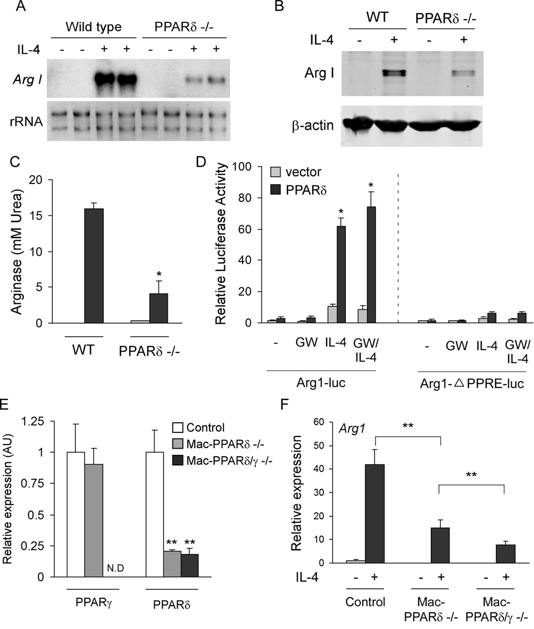
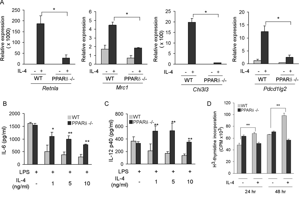
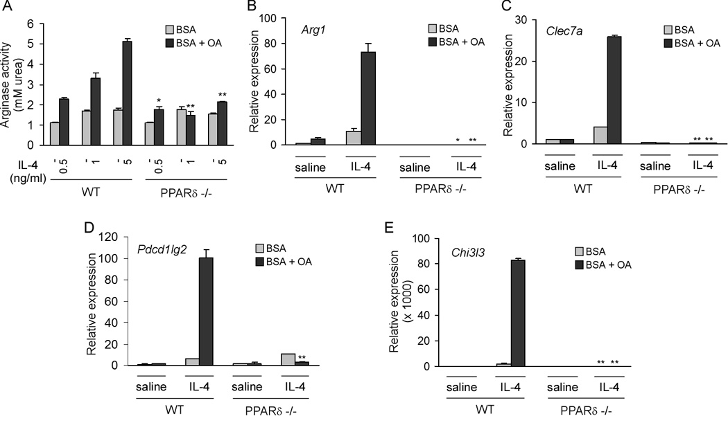
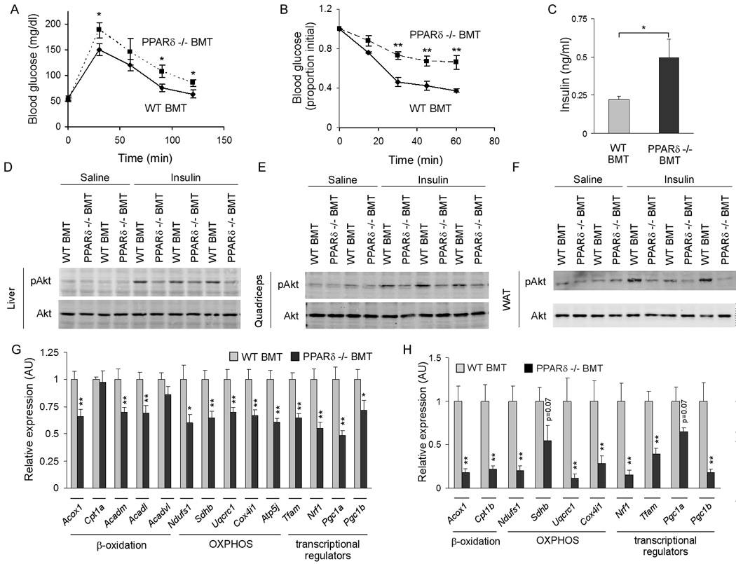
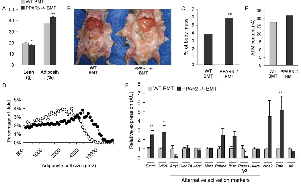
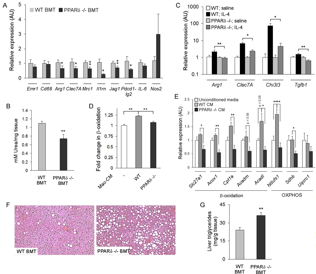
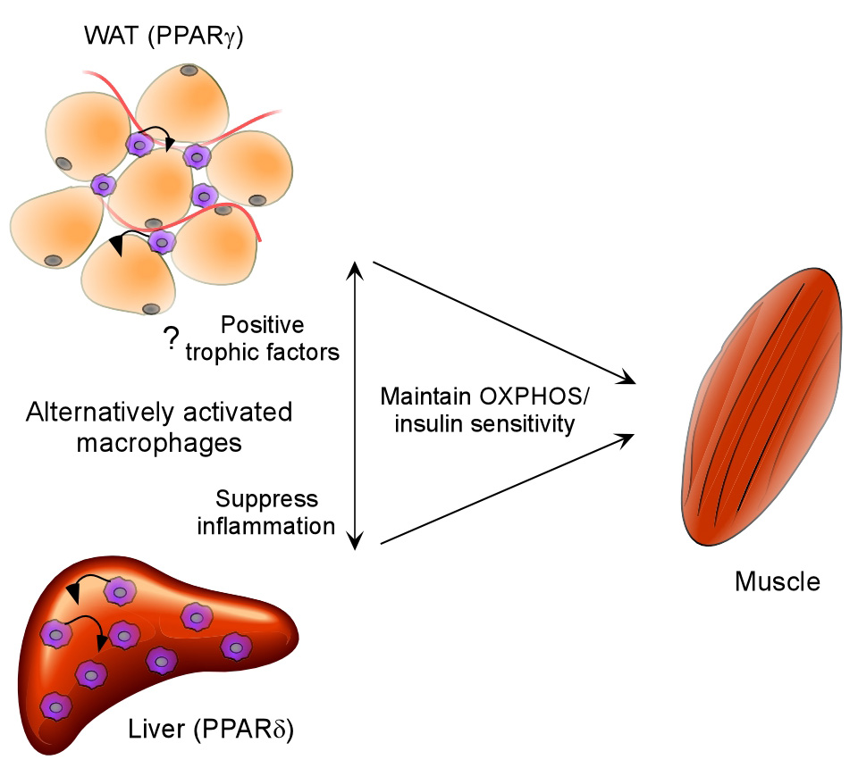
Macrophage infiltration and activation in metabolic tissues underlie obesity-induced insulin resistance and type 2 diabetes. While inflammatory activation of resident hepatic macrophages potentiates insulin resistance, the functions of alternatively activated Kupffer cells in metabolic disease remain unknown. Here we show that in response to the Th2 cytokine interleukin-4 (IL-4), peroxisome proliferator-activated receptor delta (PPARdelta) directs expression of the alternative phenotype in Kupffer cells and adipose tissue macrophages of lean mice. However, adoptive transfer of PPARdelta(-/-) (Ppard(-/-)) bone marrow into wild-type mice diminishes alternative activation of hepatic macrophages, causing hepatic dysfunction and systemic insulin resistance. Suppression of hepatic oxidative metabolism is recapitulated by treatment of primary hepatocytes with conditioned medium from PPARdelta(-/-) macrophages, indicating direct involvement of Kupffer cells in liver lipid metabolism. Taken together, these data suggest an unexpected beneficial role for alternatively activated Kupffer cells in metabolic syndrome and type 2 diabetes.


Figures

Figure 1
Figure 1. PPARδ regulates expression of arginase I in alternatively activated macrophages
(A–C) Decreased levels of arginase I mRNA (A), protein (B), and enzymatic activity (C) in IL-4-stimulated PPARδ −/−BMDMs. (D) Activation of the arginase I promoter by PPARδ/RXR heterodimers and IL-4. Mutation of PPAR response element in the arginase I enhancer abolishes transcriptional activation by PPARδ and IL-4. (E–F) Attenuated induction of arginase I in PPARδ/γ double knockout macrophages. (E) Real time analysis of PPARδ and PPARγ expression in BMDMs generated from control, macrophage-specific PPARδ null and macrophage-specific PPARδ/γ null mice. Primers specific for exon 4 of PPARδ or exon 1 of PPARγ were used to quantify excision efficiency in BMDMs. (F) qRT-PCR analysis of arginase I mRNA in various genotypes. Data presented as mean ± s.e.m. *P < 0.05, **P<0.01.
Figure 2
Figure 2. PPARδ is required for expression of the immune phenotype of alternative macrophage activation
(A) Decreased expression of markers of alternative activation in IL-4-stimulated PPARδ −/− BMDM. Relative expression of alternative activation mRNAs was quantified using by qRT-PCR. Retnla, resistin-like a; Mrc1, mannose receptor; Chi3l3, chitinase 3-like 3; Pdcd1lg2, programmed cell death 1 ligand 2. (B, C) PPARδ is required for suppression of IL-6 and IL-12 production in alternatively activated BMDMs. (D) PPARδ is required for the mitogenic response to IL-4 in BMDMs. Data presented as mean ± s.e.m. *P < 0.05, **P < 0.01.
Figure 3
Figure 3. Conjugated oleic acid potentiates alternative macrophage activation
(A) Oleic acid potentiates IL-4 induction of arginase activity in wild type, but not PPARδ−/− BMDM. (B–E) PPARδ is required for potentiation of alternative activation by oleic acid. BMDMs from 129/SvJ wild type and PPARδ null mice were stimulated IL-4 for 48 hours in the presence of BSA or BSA:oleic acid conjugate. qRT-PCR analysis were performed on mRNAs for Arg1 (arginase I), Clec7a (dectin-1), Pdcd1lg2 (programmed cell death 1 ligand 2), and Chi3l3 (chitinase 3-like 3). Data presented as mean ± s.e.m. *P < 0.05, **P < 0.01.
Figure 4
Figure 4. Hematopoietic deficiency of PPARδ exacerbates insulin resistance and impairs glucose tolerance
(A) Oral glucose tolerance test (1g/kg) were carried out in male WT BMT and PPARδ −/− BMT mice after 18 weeks of high fat diet (n=5–7 per cohort). (B) Insulin tolerance test (0.65U/kg) were performed in obese mice after a 4 hour fast (n=5–7 per cohort). Similar results were obtained in two other cohorts of transplanted mice (n=7 per group). (C) Fasting serum levels of insulin in WT BMT and PPARδ −/− BMT mice after a 5 hr fast. (D–F) Impairment in insulin action in PPARδ −/− BMT mice. Total cell lysates were immunoblotted for pAkt or total Akt in liver (D), quadriceps muscle (E) and epididymal WAT (F). (G, H) Mitochondrial dysfunction in peripheral tissues of PPARδ −/− BMT mice. Relative transcript levels for genes encoding key enzymes in β-oxidation, oxidative phosphorylation (OXPHOS), and of transcriptional regulators controlling these pathways in liver (G) and quadriceps (H). Data presented as mean ± s.e.m. *P < 0.05, **P < 0.01.
Figure 5
Figure 5. Increased adiposity in PPARδ −/− BMT mice
(A) Body composition was quantified by dual-energy X-ray absorptiometry in weight matched transplanted mice (n=7 per cohort). (B) Representative images of necropsied WT BMT and PPARδ null BMT mice after 22 weeks of high fat diet. (C) Increased epididymal fat pad mass in PPARδ null BMT mice (n=5 per cohort). (D) Increased adipocyte cell size in PPARδ null BMT mice. Adipocyte cell size was measured using dark field images. (E, F) Macrophage content and activation in white adipose tissue. ATM content was determined by immunostaining for the macrophage antigen F4/80, paired T-test P value = 0.16; n=4 per cohort (E). (F) Interrogation of ATM activation state by qRT-PCR. Emr1, F4/80; Cd68, macrosialin; Arg1, arginase I; Clec7A, dectin-1; Jag1, jagged 1; Mrc1, mannose receptor; Retlna, resistin-like alpha; Il1rn, IL-1 receptor antagonist; Pdcd1lg2, programmed cell death 1 ligand 2; Il4ra, IL-4 receptor alpha; Nos2, inducible nitric oxide synthase; Il6, interleukin-6; Tnfa, tumor necrosis factor alpha. Data presented as mean ± s.e.m. *P < 0.05, **P < 0.01.
Figure 6
Figure 6. Impaired alternative activation of Kupffer cells and hepatic dysfunction in PPARδ−/− BMT mice
(A) Decreased expression of signature genes for alternative activation macrophages in PPARδ −/− BMT livers (n=5–7 per cohort). (B) Reduction in liver arginase activity in PPARδ−/− BMT mice (n=5–7 per cohort). (C) IL-4 is unable to induce alternative activation of Kupffer cells in PPARδ −/− mice (n=4 per cohort). Arg1, arginase I; Clec7a, dectin-1; Chi3l3, chitinase 3-like 3; Tgfb1, (transforming growth factor β1). (D, E) Treatment of primary hepatocytes with macrophage conditioned media alters their oxidative metabolism. Conditioned media from PPARδ null macrophages suppresses oxidative metabolism, as monitored by β-oxidation of fatty acids (D) and expression of fatty acid oxidation and OXPHOS genes (E). (F, G) Histologic (F) and biochemical (G) evidence of increased triglyceride accumulation in livers of PPARδ −/− BMT mice. Data presented as mean ± s.e.m. *P < 0.05, **P < 0.01.
Figure 7
Figure 7
Model highlighting the metabolic functions of alternatively activated macrophages. PPARγ transcriptional signaling is required for maturation of these cells in adipose tissue, whereas PPARδ controls the expression of alternative phenotype in Kupffer cells of obese mice. Two potential mechanisms by which alternatively activated macrophages improve insulin action in obese mice are: paracrine inhibition of inflammation and secretion of trophic factors that can directly modulate oxidative metabolism in parenchymal cells (small arrows).

Comment in

References

    1. Arkan MC, Hevener AL, Greten FR, Maeda S, Li ZW, Long JM, Wynshaw-Boris A, Poli G, Olefsky J, Karin M. IKK-beta links inflammation to obesity-induced insulin resistance. Nat Med. 2005;11:191–198. - PubMed
    1. Barak Y, Liao D, He W, Ong ES, Nelson MC, Olefsky JM, Boland R, Evans RM. Effects of peroxisome proliferator-activated receptor delta on placentation, adiposity, and colorectal cancer. Proc Natl Acad Sci U S A. 2002;99:303–308. - PMC - PubMed
    1. Bouhlel MA, Derudas B, Rigamonti E, Dievart R, Brozek J, Haulon S, Zawadzki C, Jude B, Torpier G, Marx N, et al. PPARgamma activation primes human monocytes into alternative M2 macrophages with anti-inflammatory properties. Cell Metab. 2007;6:137–143. - PubMed
    1. Cai D, Yuan M, Frantz DF, Melendez PA, Hansen L, Lee J, Shoelson SE. Local and systemic insulin resistance resulting from hepatic activation of IKK-beta and NF-kappaB. Nat Med. 2005;11:183–190. - PMC - PubMed
    1. Chawla A, Lee C, Barak Y, He W, Rosenfeld J, Liao D, Han J, Kang H, Evans R. PPAR delta is a very low-density lipoprotein sensor in macrophages. Proc Natl Acad Sci U S A. 2003;100:1268–1273. - PMC - PubMed

Publication types