. 2008 Jun 5;2(6):553-65.
doi: 10.1016/j.stem.2008.03.020.

Neonatal chimerization with human glial progenitor cells can both remyelinate and rescue the otherwise lethally hypomyelinated shiverer mouse

Affiliations

Neonatal chimerization with human glial progenitor cells can both remyelinate and rescue the otherwise lethally hypomyelinated shiverer mouse

Martha S Windrem et al. Cell Stem Cell. .

Abstract

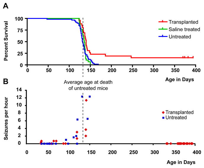
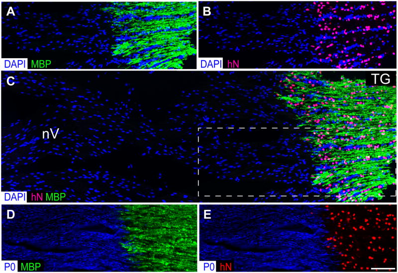
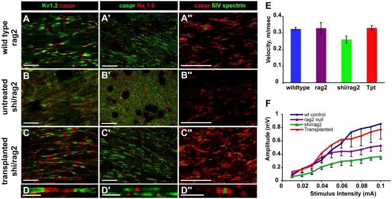
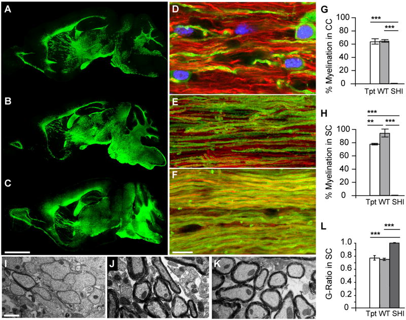
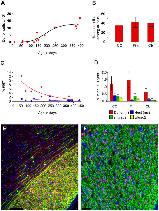
Congenitally hypomyelinated shiverer mice fail to generate compact myelin and die by 18-21 weeks of age. Using multifocal anterior and posterior fossa delivery of sorted fetal human glial progenitor cells into neonatal shiverer x rag2(-/-) mice, we achieved whole neuraxis myelination of the engrafted hosts, which in a significant fraction of cases rescued this otherwise lethal phenotype. The transplanted mice exhibited greatly prolonged survival with progressive resolution of their neurological deficits. Substantial myelination in multiple regions was accompanied by the acquisition of normal nodes of Ranvier and transcallosal conduction velocities, ultrastructurally normal and complete myelination of most axons, and a restoration of a substantially normal neurological phenotype. Notably, the resultant mice were cerebral chimeras, with murine gray matter but a predominantly human white matter glial composition. These data demonstrate that the neonatal transplantation of human glial progenitor cells can effectively treat disorders of congenital and perinatal hypomyelination.


Figures

Figure 1
Figure 1. Engrafted shiverer mice exhibited substantially prolonged survival
A, Shiverer/rag2−/− mice, either engrafted with human glial progenitor cells (GPCs) at birth (n=26, red), injected with saline (n=29, green), or untreated (n=59, blue) were maintained in small group housing and monitored daily until death. The Kaplan-Meier survival graph, plotting the percentage of each group alive as a function of age in weeks, shows that most mice die between 18 and 20 weeks. However, a fraction of engrafted mice (n=6, or 23.1%) lived substantially longer than any control mouse; 4 survived more than one year, at which point the experiment was terminated. B, Shiverer mice uniformly manifested a seizure disorder that was typically apparent by 5 weeks of age, and then worsened between 16–18 weeks. When seizure frequency was scored by video with blinded post hoc assessment, both transplanted and control shiverers were noted to seize frequently during weeks 18–20, corresponding to the time span during which most mice died. However, the seizure incidence among the transplanted shiverers fell thereafter, such that by 47 weeks of age, all surviving mice were seizure-free.
Figure 2
Figure 2. Perinatal grafts of human glial progenitors yield widespread and dense host myelination
A–B. Serial sagittal images of an engrafted shi/shi x rag2−/− brain, sacrificed at 1 year of age. Each image in a and b represents a montage of 50–100 images at 10×. Each series begins 750 μm lateral to the midline, and continues at 600 μm intervals. A. human donor cells, immunolabeled in 14 μm cryosections using an anti-human nuclear antibody (hN; red). B. Alexa 488-labeled myelin basic protein (MBP; green) in sections adjacent or nearly so to their matched sections in A. All major white matter tracts, including those of the corpus callosum, capsules, striatum, fimbria, cerebellum and brainstem heavily express MBP. C–G. Black-and-white images of MBP-immunoreactive fibers in a number of sites reveal high efficiency axonal myelination; all images of transplanted shi/shi x rag2−/− mice at > 1 year post-transplant. C, The rostral striatum, corpus callosum, and neocortical layers 5 and 6 are shown in sagittal section. D. Higher magnification of c shows the MBP-defined myelination of individual fibers within the striatum, as well as the larger bundles of corticostriatal and striopallidal fibers. E. Donor-myelinated MBP+ fibers in a longitudinal section of the cervical spinal cord; dorsal column to the left, central gray to the right. F. Interwoven donor-myelinated fibers of the brainstem, in the pontine base. G. Donor-derived MBP in the conus medullaris; exiting myelinated roots of the cauda equina to the left. Scale: A, B = 2.5 mm; C = 200 μm; D = 40 μm; E = 50 μm; F = 60 μm; G = 125 μm
Figure 3
Figure 3. Transplanted GPCs invade the cranial and spinal roots but obey the CNS-PNS border
Transplanted mice exhibited robust myelination throughout the entire CNS neuraxis by 9 months of age, that included not only the brain, brainstem, cerebellum and spinal cord, but also the cranial nerves, ganglia, and both cranial and spinal roots (see Figs. 2 and 4). Of note, the invasion of human glial progenitor cells, all derived from the fetal forebrain, was sharply delimited to the CNS, with no invasion whatsoever of the peripheral nerves beyond the root take-offs. A–B show both the dense concentration of human donor cells (anti-human nuclear antigen, red) in the trigeminal ganglion (TG), and the concurrent prohibition of donor cell infiltration into the trigeminal nerve (nV), a peripheral nerve. Accordingly, donor-derived myelin (MBP, green) was limited to the ganglion and trigeminal nerve take-off, and did not extend into nV proper. C shows a wider field color composite of A–B (A–B correspond to the boxed area of C), further demonstrating that the transplanted GPCs strictly respect the CNS-PNS border. In contrast, D–E show an adjacent section stained for the peripheral myelin protein P0 (blue), and for either human nuclear antigen (red) or central myelin basic protein (green). The human cells are seen to have stopped at the P0 protein-defined threshold to the PNS. Scale=50 μm.
Figure 4
Figure 4. Engrafted shi/shi brains exhibit restored nodes of Ranvier and callosal conduction velocities
The expression patterns of several antigens characteristic of nodes of Ranvier, including nodal (NaV1.6, βIV-spectrin), paranodal (Caspr) and juxtaparanodal (KV1.2) proteins, were investigated in the spinal cord (A–D and A″–D″) and optic nerves (A″–D″) of normally-myelinated wild-type (rag2−/−) mice (A–A″), and compared to the corresponding expression patterns in the optic nerves of both untreated (B–B″), and transplanted (C–C″ and D–D″) shiverer × rag2−/− mice. The nodal architecture of the transplanted shiverers was indistinguishable from that of wild-type controls for every antigen tested; both exhibited the sodium channel and spectrin clustering, flanked by the paranodal Caspr and juxtaparanodal Kv1.2, of mature nodes. The nodal integrity of the transplanted shiverers (C–C″ and D–D″) contrasted sharply to the disorganized and indistinct antigen expression patterns of the untreated mice (B–B″), in which neither nodal channel clustering nor paranodal Caspr sequestration was noted. E–F. Transcallosal responses were evoked by electrical stimulation in mice in vivo. E, plots the transcallosal conduction velocities obtained from wild-type, rag2−/−, shiverer x rag2−/−, and transplanted shiverer x rag2−/− mice, all assessed between 12–13 months after neonatal transplant. F, graphs the telationship between stimulus intensity and signal amplitude in C3H wild-type mice, rag2−/− mice, shiverer x rag2−/− mice, and transplanted shiverer x rag2−/− mice, respectively.
Figure 5
Figure 5. Myelination and axonal ensheathment were progressive over time
A–C, Sagittal sections of hGPC-implanted mice immunolabeled for MBP (green) at 20 weeks (A), 35 weeks (B) and 52 weeks (C). A, Major white matter regions of the brain, including the corpus callosum, fimbria, optic tract, and both the cerebral and cerebellar peduncles, are already myelinated at 20 weeks. B. At 35 weeks, the area of dense myelination has expanded into the midbrain and hindbrain. C. By a year, myelin was well-distributed, and myelination appeared complete, throughout the forebrain and hindbrain, and includes the lowers layers of neocortex, the colliculi, the pons and medulla, as well as the major corticopontine and corticospinal tracts. D–F, corresponding confocal optical sections of transplanted shiverer mouse corpus callosum taken at 20 (D), 35 (E) and 52 (F) weeks, immunolabeled for neurofilament (red) and MBP (green), reveal the progressive increase in axonal ensheathment with time. G–H, plot the proportion of MBP-ensheathed axons, as determined by confocal analysis, in the corpus callosa (CC) (G) and cervical corticospinal tracts of the spinal cord (SC) (H) in 1 year-old transplanted shi/shi × rag2−/− mice compared to both untreated and wild-type rag2−/− controls. At both sites, most axons in the transplant recipients were ensheathed by MBP-defined myelin. In contrast, no ensheathment was noted in their untreated counterparts. I–K, electron micrographs of spinal cord cross-sections in untreated shi/shi × rag2−/− mice (I), wild-type rag2−/− controls (J), and 1-year old transplanted shi/shi × rag2−/− (K). L plots the G-ratios calculated in 1 year-old implanted shiverers, compared to their wild-type and untreated shiverer controls. The correlation between myelin sheath thickness and axonal diameter in implanted mice is indistinguishable from that of wt/rag2 null mice, while myelin sheaths are virtually undetectable in untreated shiverers. Scale: A–C, 2.5 mm; D–F, 10 μm; I–K, 1 μm
Figure 6
Figure 6. Long-term survival was associated with humanization of the recipient white matter
A. Perinatally transplanted human GPCs increase in number asymptotically over the course of a year. From an initial dose of 300,000 on postnatal day 1, the cells increase to an average of 12 million/brain by one year post transplantation (y = −9,898,000 + 733,632×+ (−5709)2; r2 = 0.83). B. By one year, donor cells comprised over 40% of all cells in the fimbria and cerebellar white matter, and over a third in the corpus callosum. Since the total cell count includes host vascular cells and microglia, the human donor-derived cells appeared to comprise a net majority of all glial cells by that stage. C. Over the year after implantation, the rate of human GPC proliferation in white matter declined exponentially (red; y = 14.013e−0.0475×; r2 = 0.79). At 8 weeks, 12.35% of human GPCs in the mouse corpus callosum are Ki67 positive, but by one year, an average of 1.22% are Ki67%. From 5 to 12 months, the percentage of Ki67+ mouse cells in the corpus callosum of untreated rag2 null mice also declines exponentially, but beginning at a lower rate (purple; y = 2.9154e−0.0497×; r2 = 0.83). The Ki67+ percentages of endogenous mouse cells in the same sections of transplanted mice from which the hGPC percentages were obtained, however, do not follow a pattern of exponential decline (blue; y = 1.3684e-0.02× r2 = 0.1855). D. At one year, the percentage of hGPCs in white matter that are Ki67+ (red) exceeds that of the endogenous mouse cells in the same mice (blue), as well as that of untreated rag2 null mice (yellow), and that of 4 month old untreated shiverer/rag2 homozygotes (green) in corpus callosum, fimbria and cerebellum. E–F. Progressive myelination (MBP, green) of mouse axons (neurofilament, in red) was attended by chimerization of the recipient white matter, such that by 20 weeks, host cells (DAPI, blue) are exceeded by human donor cells (human nuclear antigen, hNA, purple, as blue co-labeled with hNA, red). E. Parasagittal section including dorsal callosum and overlying cortex of a transplanted shiverer at 20 weeks, showing human donor-derived myelination of callosum, and admixture of host (blue, DAPI) and donor cells (purple, as blue co-labeled with hNA, red). Both myelinated (MBP, green) and unmyelinated (NF, far red) fibers are evident traversing lower cortical layers. F. Higher magnification section through fimbria of hippocampus, showing myelinated fibers viewed en face, with admixed mouse (blue) and human (purple, representing blue co-labeled with hNA, red) cells. Scale: E, 100 μm; F, 50 μm.

Comment in

References

    1. Archer D, Cuddon P, Lipsitz D, Duncan I. Myelination of the canine central nervous sytem by glial cell transplantation: A model for repair of human myelin disease. Nature Medicine. 1997;3:54–59. - PubMed
    1. Archer DR, Leven S, Duncan ID. Myelination by cryopreserved xenografts and allografts in the myelin-deficient rat. Exp Neurol. 1994;125:268–277. - PubMed
    1. Back SA, Luo NL, Borenstein NS, Levine JM, Volpe JJ, Kinney HC. Late oligodendrocyte progenitors coincide with the developmental window of vulnerability for human perinatal white matter injury. J Neurosci. 2001;21:1302–1312. - PMC - PubMed
    1. Deguchi K, Oguchi K, Takashima S. Characteristic neuropathology of leukomalacia in extremely low birth weight infants. Pediatr Neurol. 1997;16:296–300. - PubMed
    1. Eftekharpour E, Karimi-Abdolrezaee S, Wang J, El Beheiry H, Morshead C, Fehlings M. Myelination of congenitally dysmyelinated spinal cord axons by adult neural precursor cells results in formation of nodes of Ranvier and improved axonal conduction. J Neuroscience. 2007;27:3416–3428. - PMC - PubMed

Publication types

MeSH terms