. 2008 Jun 10;105(23):8014-9.
doi: 10.1073/pnas.0802874105. Epub 2008 Jun 3.

Yeast UCS proteins promote actomyosin interactions and limit myosin turnover in cells

Affiliations

Yeast UCS proteins promote actomyosin interactions and limit myosin turnover in cells

Matthew Lord et al. Proc Natl Acad Sci U S A. .

Abstract

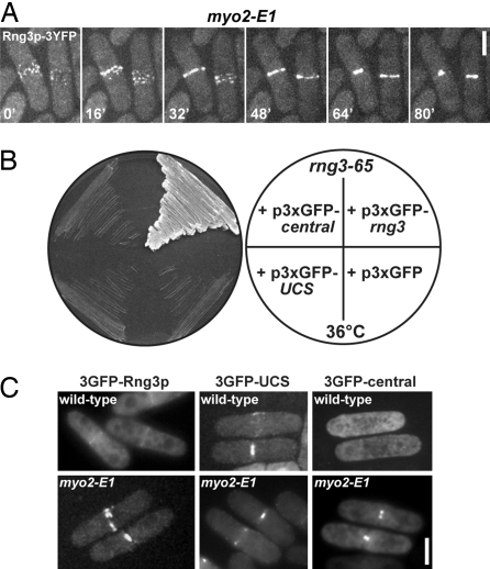
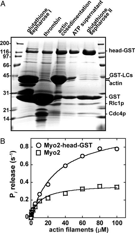
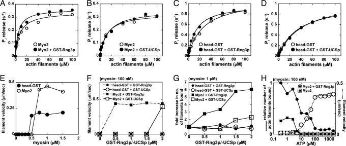
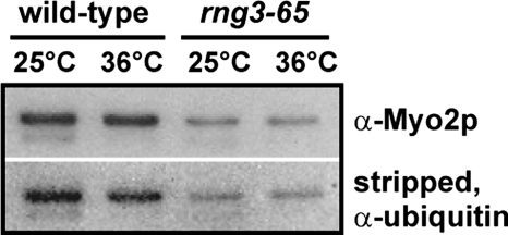
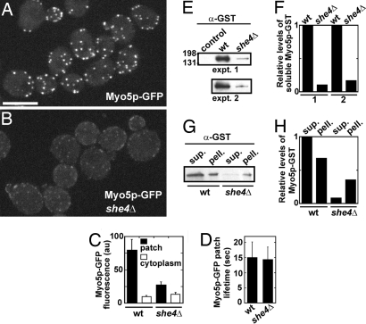
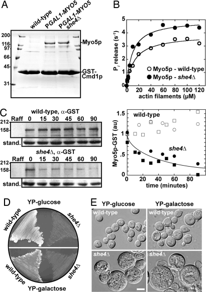
Two functions are proposed for the conserved family of UCS proteins: helping to fold myosin motor proteins and stimulating the motor function of folded myosins. We examined both functions in yeast. The fission yeast UCS protein (Rng3p) concentrates in nodes containing myosin-II (Myo2) and other proteins that condense into the cytokinetic contractile ring. Both the N-terminal (central) and C-terminal (UCS) domains of Rng3p can concentrate independently in contractile rings, but only full-length Rng3p supports contractile ring function in vivo. The presence of Rng3p in ATPase assays doubles the apparent affinity (K(ATPase)) of both native Myo2 and recombinant heads of Myo2 for actin filaments. Rng3p promotes gliding of actin filaments by full-length Myo2 molecules, but not Myo2 heads alone. Myo2 isolated from mutant strains defective for Rng3p function is soluble and supports actin filament gliding. In budding yeast the single UCS protein (She4p) acts on both myosin-I isoforms (Myo3p and Myo5p) and one of two myosin-V isoforms (Myo4p). Myo5p turns over approximately 10 times faster in she4Delta cells than wild-type cells, reducing the level of Myo5p in cells 10-fold and in cortical actin patches approximately 4-fold. Nevertheless, Myo5p isolated from she4Delta cells has wild-type ATPase and motility activities. Thus, a fraction of this yeast myosin can fold de novo in the absence of UCS proteins, but UCS proteins promote myosin stability and interactions with actin.


Conflict of interest statement

The authors declare no conflict of interest.

Figures

Fig. 1.
Fig. 1.
Rng3p central and UCS domains can independently target the contractile ring but both are needed for Rng3p function in vivo. (A) Spinning disk confocal fluorescence micrographs of live myo2-E1 cells expressing Rng3p-3YFP (MLP 683) at 23°C. Cells were mounted on 25% gelatin pads in EMM medium. A time-lapse series of two cells captured at 16-min intervals is shown. Stacks of 10 confocal z sections at intervals of 0.5 μm were collected (projected at maximum intensity) at each time point. (Scale bar: 5 μm.) (B) Viability of a rng3-65 strain (MLP 570) carrying p3xGFP-rng3 (positive control), empty vector (negative control), p3xGFP-UCS, and p3xGFP-central. Vectors contained the weak-strength 81nmt1 inducible promoter. Transformants were streaked on an EMM Ura plate and grown at 36°C. (C) Epifluorescence images of live wild-type (MLP 479) (Upper) and myo2-E1 (TP 60) (Lower) cells containing plasmids expressing 3GFP-Rng3p (p3xGFP-rng3), 3GFP-UCS (p3xGFP-UCS), or 3GFP-central (p3xGFP-central). Cells were mounted in EMM Ura medium and photographed after growth in EMM Ura medium at 25°C. (Scale bar: 5 μm.)
Fig. 2.
Fig. 2.
Purification of active Myo2-head-GST. (A) Coomassie-stained SDS/PAGE gel summarizing the purification steps of head-GST. Outside lanes display molecular mass markers (kDa). Representative head-GST samples taken during the purification are indicated (left to right): head-GST affinity purified by enrichment of GST-tagged light chains/head on glutathione-Sepharose; thrombin-treated head (removes GST from light chains); head-GST pelleted by cosedimentation with actin filaments in the absence of ATP; soluble head-GST recovered by resuspending the actin-head-GST pellet in the presence of ATP followed by a second round of actin filament pelleting; purified head-GST after removal of residual actin by rebinding of head-GST to glutathione Sepharose. (B) Head-GST (circles) and Myo2 (squares) actin-activated Mg2+-ATPase activity shown as a function of polymerized actin concentration. Basal head-GST and Myo2 ATPase activities generated from control reactions (lacking actin filaments) were subtracted from all rates derived in the presence of polymerized actin (0.5–100 μM based on the concentration of polymerized actin subunits). All assays included myosin at a final concentration of 200 nM in 2 mM Tris·HCl (pH 7.2), 10 mM imidazole, 100 mM KCl, 0.1 mM CaCl2, 3 mM MgCl2, 2 mM ATP, and 1 mM DTT. Each curve was generated from average values obtained from five different datasets and fit to Michaelis–Menten kinetics, using KaleidaGraph software.
Fig. 3.
Fig. 3.
Full-length Rng3p and Myo2p are required for maximal motor activity in vitro. (A–D) Actin-activated Mg2+-ATPase activity shown as a function of polymerized actin concentration for Myo2 plus/minus GST-Rng3p (A), Myo2 plus/minus GST-UCS (B), Head-GST plus/minus GST-Rng3p (C), and Head-GST plus/minus GST-UCS (D). Basal ATPase activities were subtracted from all rates derived in the presence of polymerized actin (0.5–100 μM). All assays included myosin at a final concentration of 200 nM (±800 nM GST-Rng3p/-UCSp) in 2 mM Tris·HCl (pH 7.2), 10 mM imidazole, 100 mM KCl, 0.1 mM CaCl2, 3 mM MgCl2, 2 mM ATP, and 1 mM DTT. For each curve, three separate datasets were averaged and displayed as a single dataset that was fit to Michaelis–Menten kinetics, using KaleidaGraph software. Activities measured in the presence of GST-Rng3p (or GST-UCS) were always recorded in unison with corresponding measurements lacking GST-Rng3p or GST-UCS. (E) The ability of head-GST and Myo2 to support binding and in vitro motility of actin filaments in 1 mM ATP is plotted as a function of the myosin concentration applied to motility chambers. (F and G) The ability of head-GST and Myo2 to support in vitro motility is plotted as a function of GST-Rng3p or GST-UCS concentration. (F) Head-GST and Myo2 were delivered into chambers at a low concentration (100 nM) that does not support filament binding and motility. (G) Head-GST and Myo2 were delivered into chambers at a high concentration (1 μM) that supports filament motility. The number of filaments bound by myosin at the cover-slip surface was averaged by using four 130-μm2 frames taken from fluorescence micrographs from two independent experiments. Average filament numbers are plotted as relative values, with 1 being equal to the number of filaments bound in the absence of GST-Rng3p or GST-UCS. (H) The relative number of actin filaments (where 1 represents the highest recorded value) bound by Myo2 were plotted as a function of the ATP concentration in the running buffer (small filled squares, 50 nM Myo2 alone; small filled circles, 50 nM Myo2 plus 500 nM GST-Rng3p). Filament numbers were quantitated and averaged as described in G. The rate at which Myo2-bound actin filaments move in these assays is also displayed as a function of ATP concentration (large open squares, 50 nM Myo2 alone; large open circles, 50 nM Myo2 plus 500 nM GST-Rng3p).
Fig. 4.
Fig. 4.
Isolation of soluble Myo2 from rng3-65 cells. Myo2p and its GST-tagged light chains were overexpressed and 1-step purified from wild-type (MLP 509) and rng3-65 (MLP 586) cells in which the myo2 promoter was replaced with the nmt41 (medium-strength) inducible promoter. Myo2 expression was induced for 22 h at 25°C followed by further induction for 4 h at 25°C or 36°C. Samples purified in one-step by affinity chromatography on glutathione-Sepharose were run on SDS/PAGE gels, transferred to nitrocellulose, and probed with anti-Myo2p tail antibodies to compare levels of Myo2p. The blot was stripped with 4% trichloroacetic acid and reprobed with anti-ubiquitin antibodies to compare levels of Myo2p ubiquitination. Samples were prepared identically and the volumes loaded on gels normalized based on the relative protein concentrations of soluble lysed cell extracts.
Fig. 5.
Fig. 5.
UCS protein She4p maintains the cellular levels of budding yeast Myo5p (myosin-I). Genomic integration at the MYO5 locus was used to generate MYO5-GFP and MYO5-GST strains. Spinning disk confocal fluorescence microscopy of live wild-type (MLY 701) and she4Δ (MLY 724) budding yeast cells expressing Myo5p-GFP was used to generate images, which were analyzed using Image J software. Cells were mounted on 25% gelatin pads in CSM medium. Still or time-lapse images were recorded in stacks of 12 confocal z sections (at intervals of 0.5 μm) and projected at maximum intensity. Relative levels of Myo5-GST recovered from wild-type (MLY 697) and she4Δ (MLY 720) cells were estimated by densitometry of Myo5-GST bands, using Image J software. Myo5p-GST bands were detected by using anti-GST antibodies after immunoblotting of SDS/PAGE gels containing glutathione Sepharose-enriched or fractionated cell lysate samples. (A and B) The subcellular localization of Myo5p-GFP (as patches) in wild-type and she4Δ cells. (Scale bar: 5 μm.) (C) Relative levels of Myo5p-GFP fluorescence detected in both cortical patches and cytoplasm of wild-type and she4Δ cells (n = 10). (D) Average lifetimes of Myo5p-GFP in cortical patches (n = 50) of wild-type and she4Δ cells. (E) Immunoblots indicating the levels of native Myo5p-GST enriched from control (JC 1284, a strain lacking integration of GST at the MYO5 locus), wild-type (MLY 697), and she4Δ (MLY 720) cells. An ≈160-kDa band reacted with α-GST antibodies reflecting the predicted size of Myo5p-GST (155 kDa). The experiment was performed in duplicate, using two alternative segregants corresponding to MLY 697 and 720. (F) Relative levels of Myo5p-GST detected from immunoblots shown in E. (G) Immunoblots indicating levels of native Myo5p-GST in wild-type and she4Δ cell extracts. Cells were lysed and spun to separate extracts into soluble supernatant (sup.) and insoluble pellet (pell.) samples. (H) Relative levels of Myo5p-GST detected from immunoblots in G.
Fig. 6.
Fig. 6.
Purification and turnover of Myo5p in the absence of She4p. The genomic MYO5 promoter was replaced with the inducible GAL1 promoter. Myo5p was co-overexpressed with calmodulin (Cmd1p) in budding yeast (A and B) or alone as a Myo5p-GST fusion (C–E). (A) Coomassie-stained SDS/PAGE gel indicating the recovery of soluble Myo5p from wild-type (control cells lacking an integrated GAL1 promoter, Y 258), GAL1promoter-MYO5 (MLY 745), and she4Δ GAL1promoter-MYO5 (MLY 758) cells. All three strains contained a plasmid (pPGAL1-GST-CMD1-TRP1 or pPGAL1-GST-CMD1-URA3) expressing GST-Cmd1p from the galactose-inducible promoter. Protein was enriched by one-step purification on glutathione Sepharose. Outside gel lanes show molecular mass markers (kDa). (B) Actin-activated ATPase activity of Myo5p plotted as a function of polymerized actin concentration. Myo5p was purified in one step (as described in A) from wild-type and she4Δ cells. The concentration of Myo5p in these impure samples was estimated by using densitometry measurements of Coomassie-stained protein bands with rabbit skeletal muscle myosin as the standard. Basal Myo5p ATPase activities were subtracted from all rates derived in the presence of polymerized actin (1–120 μM). All assays included Myo5p at a final concentration of 50–75 nM in 2 mM Tris·HCl (pH 7.2), 10 mM imidazole, 100 mM KCl, 0.1 mM CaCl2, 3 mM MgCl2, 2 mM ATP, and 1 mM DTT. For each curve, two separate datasets were averaged and displayed as a single dataset fit to Michaelis–Menten kinetics, using KaleidaGraph software. (C) Pulse–chase experiment on the biosynthesis and turnover of Myo5p-GST. Wild-type (MLY 801, PGAL1-MYO5-GST) and she4Δ (MLY 802, she4Δ PGAL1-MYO5-GST) cells were grown in rich media containing 1% raffinose before induction of Myo5p-GST expression by addition of 2% galactose for 1 h. Cells were harvested and Myo5p-GST expression repressed by growth in rich media containing 2% glucose. Cell samples were taken before induction (Raff), after induction for 1 h (0) and at time points 15–90 min after initiating repression. Equal amounts of soluble protein in total cell extracts were separated by SDS/PAGE and analyzed by immunoblotting with antibodies to GST. The 155-kDa band is the size expected for Myo5p-GST. The graph shows the time course of the density of the Myo5p-GST bands from two experiments normalized for the intensity of the band in wild-type cells at chase time 0. Circles denote values derived from the blots on the left, squares represent data from an independent experiment. The curved line is an exponential with a half time of 26 min. (D) Wild-type (MLY 801, PGAL1-MYO5-GST) and she4Δ (MLY 802, she4Δ PGAL1-MYO5-GST) cells were grown at 36°C on rich media containing glucose (repressing conditions, Upper) and rich media containing galactose to induce overexpression of Myo5p-GST (Lower). (E) Representative DIC images of wild-type and temperature-sensitive she4Δ cells (taken from plates shown in D).

References

    1. Hutagalung AH, Landsverk ML, Price MG, Epstein HF. The UCS family of myosin chaperones. J Cell Sci. 2002;115:3983–3990. - PubMed
    1. Barral JM, Bauer CC, Ortiz I, Epstein HF. Unc-45 mutations in Caenorhabditis elegans implicate a CRO1/She4p-like domain in myosin assembly. J Cell Biol. 1998;143:1215–1225. - PMC - PubMed
    1. Kachur T, Ao W, Berger J, Pilgrim D. Maternal UNC-45 is involved in cytokinesis and colocalizes with non-muscle myosin in the early Caenorhabditis elegans embryo. J Cell Sci. 2004;117:5313–5321. - PubMed
    1. Landsverk ML, et al. The UNC-45 chaperone mediates sarcomere assembly through myosin degradation in Caenorhabditis elegans. J Cell Biol. 2007;177:205–210. - PMC - PubMed
    1. Barral JM, Hutagalung AH, Brinker A, Hartl FU, Epstein HF. Role of the myosin assembly protein UNC-45 as a molecular chaperone for myosin. Science. 2002;295:669–671. - PubMed

Publication types

MeSH terms

LinkOut - more resources