. 2008 Jun 10;105(23):8067-72.
doi: 10.1073/pnas.0709684105. Epub 2008 Jun 3.

ATP is released by monocytes stimulated with pathogen-sensing receptor ligands and induces IL-1beta and IL-18 secretion in an autocrine way

Affiliations

ATP is released by monocytes stimulated with pathogen-sensing receptor ligands and induces IL-1beta and IL-18 secretion in an autocrine way

Alessandra Piccini et al. Proc Natl Acad Sci U S A. .

Abstract

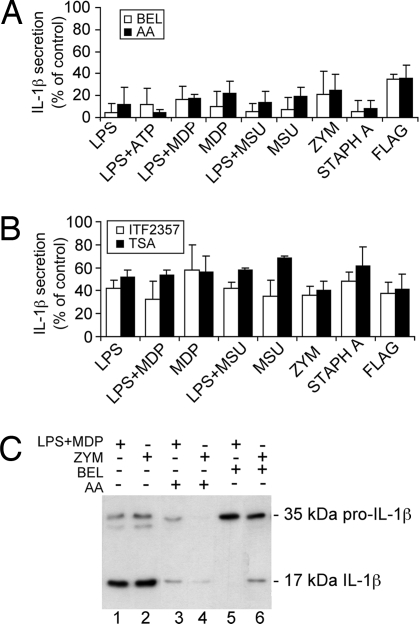
IL-1beta and IL-18 are crucial mediators of inflammation, and a defective control of their release may cause serious diseases. Yet, the mechanisms regulating IL-1beta and IL-18 secretion are partially undefined. Both cytokines are produced as inactive cytoplasmic precursors. Processing to the active form is mediated by caspase-1, which is in turn activated by the multiprotein complex inflammasome. Here, we show that in primary human monocytes microbial components acting on different pathogen-sensing receptors and the danger-associated molecule uric acid are all competent to induce maturation and secretion of IL-1beta and IL-18 through a process that involves as a first event the extracellular release of endogenous ATP. ATP release is followed by autocrine stimulation of the purinergic receptors P2X(7). Indeed, antagonists of the P2X(7) receptor (P2X(7)R), or treatment with apyrase, prevent IL-1beta and IL-18 maturation and secretion triggered by the different stimuli. At variance, blocking P2X(7)R activity has no effects on IL-1beta secretion by monocytes carrying a mutated inflammasome that does not require exogenous ATP for activation. P2X(7)R engagement is followed by K+ efflux and activation of phospholipase A(2). Both events are required for processing and secretion induced by all of the stimuli. Thus, stimuli acting on different pathogen-sensing receptors converge on a common pathway where ATP externalization is the first step in the cascade of events leading to inflammasome activation and IL-1beta and IL-18 secretion.


Conflict of interest statement

The authors declare no conflict of interest.

Figures

Fig. 1.
Fig. 1.
Kinetics of production and secretion of IL-1β in response to different PAMPs and DAMPs. Monocytes were cultured for different periods of time in the absence (CTR) or presence of LPS, MDP, zymosan (ZYM), S. aureus (STAPH A), flagellin (FLAG), or MSU, alone or in association as indicated. (A) Western blot of IL-1β in cell lysates at 3 or 15 h of exposure to stimuli. Migration of the 35-kDa pro-IL-1β molecular form is indicated. (B and C) ELISA of IL-1β released by monocytes cultured for 3, 6, or 15 h with the same PAMPs or DAMPs as in A. Note that LPS+ATP represents the amount of IL-1β released during 15 min of exposure of LPS-treated monocytes to exogenous ATP. (D) ELISA of IL-1β released by monocytes cultured for 6 h with PAMPs or DAMPs (empty columns) followed by 15 min of ATP (filled columns). Results are expressed as ng/ml/106 cells. Values are the mean ± SEM of five experiments performed on monocytes from different donors.
Fig. 2.
Fig. 2.
Inhibitors of PLA2 and HDAC prevent IL-1β secretion. (A and B) Monocytes were stimulated with the different PAMPs or DAMPs for 3 h in the absence or presence of the PLA2 inhibitors BEL (10 μM; empty columns) or AACOCF3 (AA, 40 μM; filled columns) (A) or for 15 h in the absence or presence of the HDAC inhibitors ITF2357 (100 nM; empty columns) or trichostatin A (TSA, 1 μM; filled columns) (B). Secreted IL-1β was quantified by ELISA. Data are expressed as percent of secretion with respect to untreated cells. Values are the mean ± SD of at least three experiments on monocytes from different donors (P < 0.001 and P < 0.01 in A and B, respectively). (C) Western blot analysis of supernatants from monocytes cultured for 3 h with LPS plus MDP (lanes 1, 3, and 5) or zymosan (ZYM; lanes 2, 4, and 6) in the absence (lanes 1 and 2) or presence of AACOCF3 (AA; lanes 3 and 4) or BEL (lanes 5 and 6). Migration of the 35-kDa pro-IL-1β and the 17-kDa mature IL-1β molecular forms is indicated.
Fig. 3.
Fig. 3.
Role of K+ efflux in IL-1β production, processing, and secretion. (A) IL-1β in cell lysates (Left) and supernatants (Right) of monocytes incubated 3 h alone (lanes 1) or with LPS (lanes 2 and 5) or with LPS plus MDP (lanes 3 and 4) in control medium (5 mM KCl; lanes 1–3) or KCl buffer (150 mM KCl; lanes 4 and 5). Migration of the 35-kDa pro-IL-1β and the 17-kDa mature IL-1β molecular forms is indicated. (B and C) Effect of high K+ buffer or free K+ buffer on IL-1β (B) and IL-8 (C) secreted by monocytes stimulated 3 h with the different PAMPs and DAMPs. Values are the mean of at least four experiments on monocytes from different healthy donors ± SD (P < 0.05).
Fig. 4.
Fig. 4.
All PAMPs and DAMPs induce endogenous ATP release by monocytes. (A) ATP levels in supernatants from monocytes cultured 3 h without (untreated) or with all of the different PAMPs or DAMPs in the absence (CTR) or presence of zVAD (40 μM) and ARL (200 μM) as indicated. Results are expressed as nM of ATP secreted per 106 cells. Values are the mean of at least four different experiments ± SD. (B) IL-1β secreted by monocytes from the same donors under the same experimental conditions, evaluated by ELISA (mean ± SD).
Fig. 5.
Fig. 5.
Role of P2X7R activation on IL-1β production, processing, and secretion. (A and C) IL-1β (A) and IL-8 (C) secreted by monocytes stimulated 3 h with the different PAMPs or DAMPs in the absence (CTR) or presence of oATP (300 μM), KN-62 (1 μM), and apyrase (2.5 units/ml) as indicated were quantified by ELISA. Values correspond to mean ± SEM of one representative experiment of five performed. IL-1β secretion is significantly inhibited by either oATP (P < 0.001) or KN-62 and apyrase (P < 0.01). (B) Aliquots of cell lysates were analyzed by Western blotting. Migration of the 35-kDa pro-IL-1β molecular forms is indicated. (D) IL-1β secreted by monocytes stimulated 3 h with LPS plus 15 min with ATP or nigericin (NIG), in the absence (CTR) or presence of oATP or KN-62 as indicated was quantified by ELISA. (E) IL-1β secreted by monocytes from one healthy control (HC) and one patient with CINCA syndrome was quantified by ELISA after 3 h of LPS stimulation or after additional 15 min with fresh medium supplemented with 1 mM ATP. oATP significantly inhibited secretion by HC monocytes (P < 0.001) but not by CINCA patient monocytes. Values correspond to four representative experiments ± SD.
Fig. 6.
Fig. 6.
Processing and secretion of IL-18 by monocytes stimulated with PAMPs or DAMPs. (A) Monocytes were cultured with the different stimuli as indicated for 3 or 15 h. Secreted IL-18 was determined by ELISA. Results are expressed as pg/ml/106 cells. Values are the mean ± SD of at least three of five experiments on monocytes from different donors. (B) Comparison of IL-18 secreted by monocytes from one representative healthy control (HC) and one patient with CINCA syndrome, cultured 3 h in the absence (CTR) or presence of LPS. Results are expressed as pg/ml per 106 cells ± SEM. (C) IL-18 secretion by monocytes cultured 3 h with the different stimuli in the presence of oATP. Data are expressed as percent of secretion with respect to untreated cells. Values represent the mean ± SD of at least three experiments on monocytes from different HC (P < 0.01), and the mean ± SEM of three experiments on monocytes from the CINCA patient. (D) Supernatants from monocytes cultured for 3 h with LPS plus MDP in the absence (lane 2) or presence of AACOCF3 (AA; lane 1) or BEL (lane 3) were analyzed by Western blotting with anti-IL-18. Migration of the 24-kDa pro-IL-18 and the 18-kDa IL-18 molecular forms is indicated.

References

    1. Kaczorowski DJ, Mollen KP, Edmonds R, Billiar TR. Early events in the recognition of danger signals after tissue injury. J Leukocyte Biol. 2008;83:546–552. - PubMed
    1. Akira S, Takeda K. Toll-like receptor signaling. Nat Rev Immunol. 2004;4:499–511. - PubMed
    1. Mitchell JA, Paul-Clark MJ, Clarke GW, McMaster SK, Cartwright N. Critical role of toll-like receptors and nucleotide oligomerization domain in the regulation of health and disease. J Endocrinol. 2007;193:323–330. - PubMed
    1. Rubartelli A, Lotze MT. Inside, outside, upside down: Damage-associated molecular-pattern molecules (DAMPs) and redox. Trends Immunol. 2007;28:429–436. - PubMed
    1. Dinarello CA. Interleukin-1, interleukin-1 receptors, and interleukin-1 receptor antagonist. Int Rev Immunol. 1998;16:457–499. - PubMed

Publication types

MeSH terms

LinkOut - more resources