. 2008 Jun 10;105(23):8136-41.
doi: 10.1073/pnas.0803849105. Epub 2008 Jun 3.

Plexin signaling selectively regulates the stereotyped pruning of corticospinal axons from visual cortex

Affiliations

Plexin signaling selectively regulates the stereotyped pruning of corticospinal axons from visual cortex

Lawrence K Low et al. Proc Natl Acad Sci U S A. .

Abstract

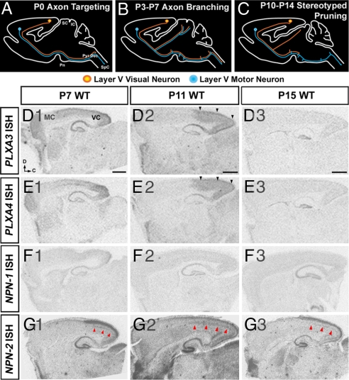
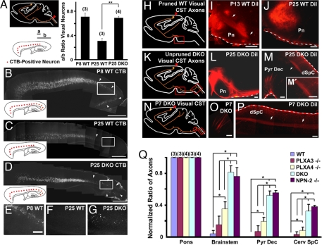
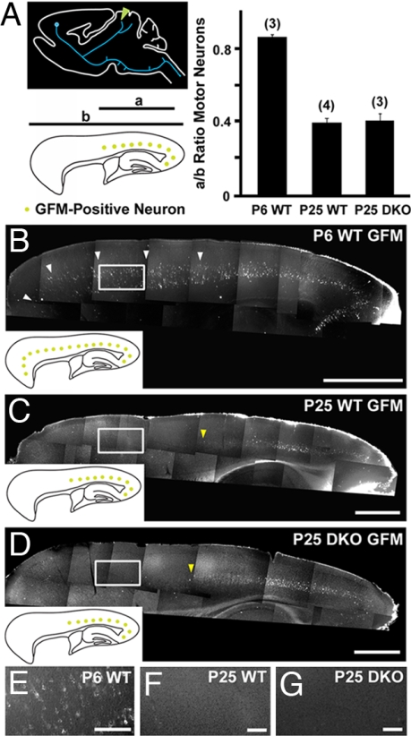
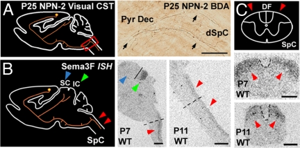
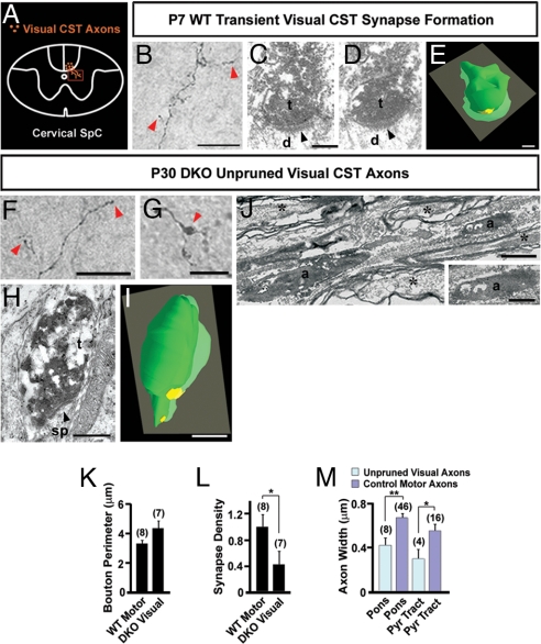
Neurons in the developing CNS tend to send out long axon collaterals to multiple target areas. For these neurons to attain specific connections, some of their axon collaterals are subsequently pruned-a process called stereotyped axon pruning. One of the most striking examples of stereotyped pruning in the CNS is the pruning of corticospinal tract (CST) axons. The long CST collaterals from layer V neurons of the visual and motor cortices are differentially pruned during development. Here we demonstrate that select plexins and neuropilins, which serve as coreceptors for semaphorins, are expressed in visual cortical neurons at the time when CST axon collaterals are stereotypically pruned. By analyzing mutant mice, we find that the pruning of visual, but not motor, CST axon collaterals depends on plexin-A3, plexin-A4, and neuropilin-2. Expression pattern study suggests that Sema3F is a candidate local cue for the pruning of visual CST axons. Using electron microscopic analysis, we also show that visual CST axon collaterals form synaptic contacts in the spinal cord before pruning and that the unpruned collaterals in adult mutant mice are unmyelinated and maintain their synaptic contacts. Our results indicate that the stereotyped pruning of the visual and motor CST axon collaterals is differentially regulated and that this specificity arises from the differential expression of plexin receptors in the cortex.


Conflict of interest statement

The authors declare no conflict of interest.

Figures

Fig. 1.
Fig. 1.
Plexin and neuropilin expression in the neocortex during CST pruning. (A–C) Diagrams of sagittal views of the brain representing different stages of CST development: axon targeting by P0, axon branching between P3 and P7, and stereotyped axon pruning between P10 and P14. (D and E) PLXA3 and PLXA4 mRNAs are expressed throughout the cortex at P7, and the expression becomes restricted to visual cortex by P11 (black arrowheads). (F) NPN-1 mRNA is not expressed in the neocortex between P7 and P15. (G) NPN-2 mRNA is expressed in the superficial and deep layers (red arrowheads) of the visual cortex between P7 and P15. D, dorsal; C, caudal; IC, inferior colliculus; MC, motor cortex; Pn, pons; Pyr Dec, pyramidal decussation; SC, superior colliculus; SpC, spinal cord; VC, visual cortex. (Scale bars: 1,000 μm.)
Fig. 2.
Fig. 2.
PLXA3 and PLXA4 are required for the stereotyped pruning of visual corticospinal axons from the spinal cord. (A–G) Layer V neurons in the visual cortex (orange) were retrogradely labeled by injecting CTB into the dorsal cervical spinal cord (red arrowhead in A). The distribution of labeled neurons in the visual cortex is normalized for comparison as diagramed in A (a/b ratio; see Methods). A Right shows that visual CST axons are not pruned from the spinal cord of P25 PLXA3/PLXA4−/− (DKO) mice (mean ± SEM; n values are indicated in parentheses). ∗∗, P < 0.01 (Student's t test). Representative images of retrogradely labeled neurons in the cortex for each set of experiments are shown in B–D. The Inset in each image summarizes the distribution of CTB-positive neurons (red) in the cortex, and the white arrowheads indicate the distribution of neurons in the visual cortex. Higher-magnification images taken from boxed regions in B–D are shown in E–G, respectively. (H–P) Anterograde tracing of axons from the visual cortex in WT and DKO mice. Red arrowheads in H, K, and N indicate the injection sites. DiI-labeled axons in WT mice are in the process of pruning at P13 (arrows in I), and at P25 axons terminate in the rostral pons (J). DiI-labeled axons in P25 DKO animals extend beyond the rostral pons (L), cross to the contralateral spinal cord at the pyramidal decussation, and terminate in the spinal cord (M). Unpruned axons in the DKO spinal cord (arrow) are shown in higher magnification in (M′). DiI-labeled axons in P7 DKO animals (arrows) grow normally into the pyramidal decussation (O) and dorsal spinal cord (P). White dashes in I, J, L, M, and P) indicate meninges and do not represent positive DiI labeling. (Q) A comparison of normalized ratios of BDA-labeled visual CST axons (mean ± SEM) present at the pons, brainstem, pyramidal decussation (Pyr Dec), and cervical spinal cord (Cerv SpC) in mice aged P30–P35 (see Methods). When compared with WT mice, significant pruning defects are found in PLXA4−/−, DKO, and NPN-2−/− mice [∗, P < 0.05 (ANOVA, Newman–Keuls test)], but not in PLXA3−/− mice. The defects in DKO and NPN-2−/− mice are also more severe than in PLXA3−/− or PLXA4−/− mice [∗, P < 0.05 (ANOVA, Newman–Keuls test)] at all levels beyond rostral pons. n values are indicated in parentheses. dSpC, dorsal spinal cord; Pn, pons. [Scale bars: 1,000 μm (B–D) and 200 μm (E–G, I, J, L, M, O, and P).]
Fig. 3.
Fig. 3.
PLXA3 and PLXA4 are not required for the stereotyped pruning of motor corticospinal axons from the superior colliculus. (A) Layer V neurons in the motor cortex (blue) were retrogradely labeled by injection of GFM in the superior colliculus (green arrowhead). The distribution of labeled neurons in the cortex is normalized for comparison as diagramed (a/b ratio; see Methods). The bar graph indicates no significant motor CST pruning defects from the superior colliculus of PLXA3/PLXA4−/− (DKO) mice (mean ± SEM; n values are indicated in parentheses). Representative images of retrogradely labeled neurons in the cortex for each set of experiments are shown in B–D. The Inset in each image summarizes the distribution of CTB-positive neurons (green) in the cortex. Layer V motor neurons that extend transient projections to the superior colliculus at P6 are indicated by white arrowheads (B). Yellow arrowheads (C and D) mark the border of GFM-positive neurons. Higher-magnification images taken from boxed regions in B–D are shown in E–G, respectively. [Scale bars: 1,000 μm (B–D) and 200 μm (E–G).]
Fig. 4.
Fig. 4.
Stereotyped pruning of the visual CST is likely to be initiated by Sema3F signaling through PLXA3 and PLXA4 and their coreceptor, NPN-2. (A) Diagram and image showing unpruned BDA-positive visual CST axons (black arrows) in the pyramidal decussation (Pyr Dec) and dorsal cervical spinal cord (dSpC) of P25 NPN-2−/− mice. (B) Sema3F mRNA expression is observed in the transient targets of visual CST axon collaterals at the inferior colliculus (green arrowheads) and dorsal spinal cord (red arrowheads) of P7 and P11 WT mice. A line is shown separating the superior and inferior colliculus. Sema3F mRNA is absent from the superior colliculus (blue arrowhead), which retains its visual CST axon collaterals. Dashed lines indicate the regions from which transverse sections in C are taken. (C) Sema3F mRNA expression in the dorsal regions of transverse sections of the spinal cord at P7 and P11 (red arrowheads). Dashed lines outline the dorsal funiculus, where CST axons are located. DF, dorsal funiculus; IC, inferior colliculus; SC, superior colliculus; SpC, spinal cord. [Scale bars: 100 μm (A), 1,000 μm (B), and 500 μm (C).]
Fig. 5.
Fig. 5.
EM analysis of visual CST axons in the spinal cord of WT and PLXA3/PLXA4−/− mice. (A) Diagram of a transverse cervical spinal cord section showing visual CST axons branching ventrally into the gray matter. The box indicates the location where the images in B, F, and G are taken. (B) A BDA-labeled transient visual CST axon in P7 WT mouse branches into the cervical spinal cord gray matter and exhibits bouton-like structures (red arrowheads). (C and D) Serial electron micrographs showing a BDA-labeled visual CST axon terminal (t) forming an asymmetric synapse (black arrowhead) with a dendrite (d) in the gray matter of the cervical spinal cord of P7 WT mouse. (E) Three-dimensional EM reconstruction of serial sections from a BDA-labeled terminal shown in C and D demonstrating an axon terminal (green) adjacent to a postsynaptic density (yellow). (F and G) Unpruned BDA-labeled visual CST axons are found in the dorsal CST of the spinal cord of P30 PLXA3/PLXA4−/− (DKO) mice. Axon branches within the gray matter contain bouton-like structures (red arrowheads). (H) An electron micrograph showing a BDA-positive visual CST axon terminal (t) in the gray matter of the spinal cord of a P30 DKO mouse. The BDA-labeled terminal contains vesicles that cluster adjacent to a postsynaptic density (black arrowhead) within a dendritic spine (sp). (I) Three-dimensional reconstruction of the axon terminal in H (green) adjacent to a postsynaptic density (yellow). (J) An electron micrograph showing unmyelinated, unpruned BDA-positive visual CST axons (a) adjacent to unlabeled myelinated axons (asterisks) within the brainstem. (K) A comparison of average bouton sizes (mean ± SEM) of adult WT motor and adult DKO visual axons in the spinal cord indicates no significant differences. (L) A comparison of average synapse number per bouton (mean ± SEM) for adult WT motor and adult DKO visual axonal boutons in the spinal cord indicates significantly fewer synapses made by unpruned adult DKO visual axonal boutons [∗, P ≤ 0.05 (Student's t test)]. (M) A comparison of average axon diameter (mean ± SEM) of BDA-positive unpruned visual axons in DKO animals and surrounding motor axons indicates that in the caudal pons and pyramidal tract (Pyr Tract) DKO unpruned visual axons are significantly smaller in size [∗, P < 0.05; ∗∗, P < 0.01 (Student's t test)]. n values are indicated in parentheses. [Scale bars: 50 μm (B), 0.25 μm (C–E), 25 μm (F and G), 0.5 μm (H, I, and Inset in J), and 1 μm (J).]

Similar articles

Cited by

References

    1. Kantor DB, Kolodkin AL. Curbing the excesses of youth: Molecular insights into axonal pruning. Neuron. 2003;38:849–852. - PubMed
    1. Low LK, Cheng HJ. A little nip and tuck: Axon refinement during development and axonal injury. Curr Opin Neurobiol. 2005;15:549–556. - PubMed
    1. Low LK, Cheng HJ. Axon pruning: An essential step underlying the developmental plasticity of neuronal connections. Philos Trans R Soc London B. 2006;361:1531–1544. - PMC - PubMed
    1. Luo L, O'Leary DD. Axon retraction and degeneration in development and disease. Annu Rev Neurosci. 2005;28:127–156. - PubMed
    1. Innocenti GM, Price DJ. Exuberance in the development of cortical networks. Nat Rev Neurosci. 2005;6:955–965. - PubMed

Publication types

MeSH terms

LinkOut - more resources