. 2008 Sep;84(3):814-23.
doi: 10.1189/jlb.0807561. Epub 2008 Jun 3.

The p110gamma isoform of phosphatidylinositol 3-kinase regulates migration of effector CD4 T lymphocytes into peripheral inflammatory sites

Affiliations

The p110gamma isoform of phosphatidylinositol 3-kinase regulates migration of effector CD4 T lymphocytes into peripheral inflammatory sites

Molly S Thomas et al. J Leukoc Biol. 2008 Sep.

Abstract

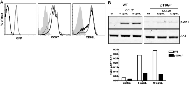
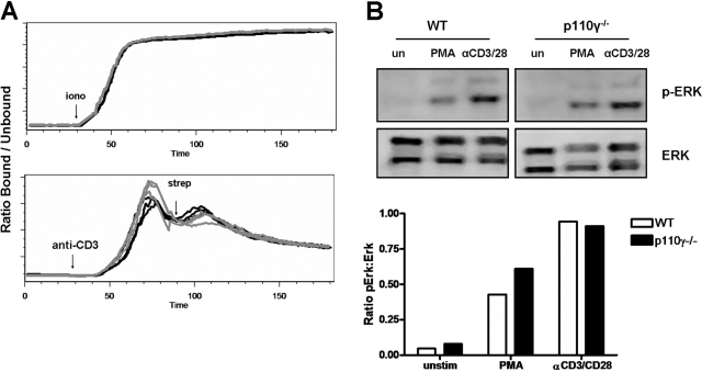
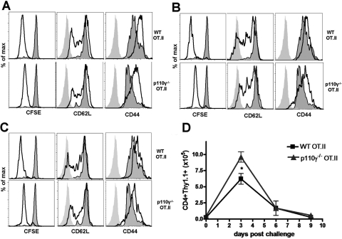
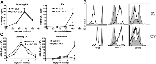
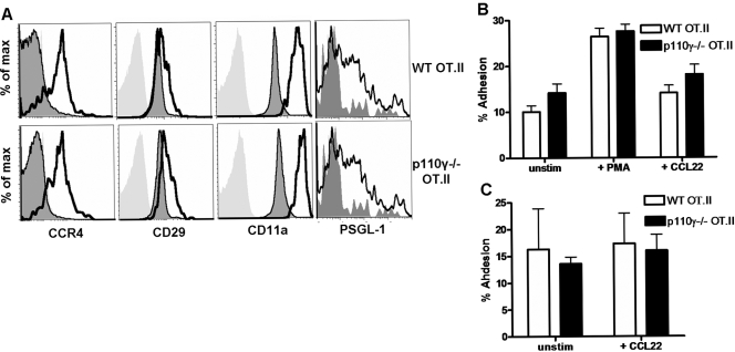
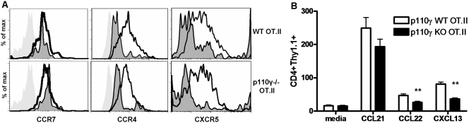
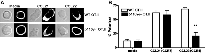
The role of PI-3K in leukocyte function has been studied extensively. However, the specific role of the p110gamma isoform of PI- 3K in CD4 T lymphocyte function has yet to be defined explicitly. In this study, we report that although p110gamma does not regulate antigen-dependent CD4 T cell activation and proliferation, it plays a crucial role in regulating CD4 effector T cell migration. Naïve p110gamma(-/-) CD4 lymphocytes are phenotypically identical to their wild-type (WT) counterparts and do not exhibit any defects in TCR-mediated calcium mobilization or Erk activation. In addition, p110gamma-deficient CD4 OT.II T cells become activated and proliferate comparably with WT cells in response to antigen in vivo. Interestingly, however, antigen-experienced, p110gamma-deficient CD4 OT.II lymphocytes exhibit dramatic defects in their ability to traffic to peripheral inflammatory sites in vivo. Although antigen-activated, p110gamma-deficient CD4 T cells express P-selectin ligand, beta2 integrin, beta1 integrin, CCR4, CXCR5, and CCR7 comparably with WT cells, they exhibit impaired F-actin polarization and migration in response to stimulation ex vivo with the CCR4 ligand CCL22. These findings suggest that p110gamma regulates the migration of antigen-experienced effector CD4 T lymphocytes into inflammatory sites during adaptive immune responses in vivo.


Figures

Fig. 1.
Fig. 1.
Phenotypic analysis of naïve, p110γ-deficient CD4 lymphocytes. (A) Naïve, p110γ-deficient lymphocytes were analyzed for GFP, CCR7, and CD62L expression by flow cytometry. Shaded histogram indicates isotype control staining, black line indicates WT, and gray line indicates p110γ−/− CD4 T cells. (B) AKT phosphorylation in naive T cells following stimulation with 5 or 10 μg/ml rmCCL21 for 1 min was analyzed by Western blotting. Densitometric analysis was preformed digitally using the Odyssey imaging system and is presented as the ratio of p-AKT to total AKT in each sample.
Fig. 2.
Fig. 2.
TCR-mediated activation of p110γ-deficient CD4 lymphocytes. (A) Calcium flux in naïve, p110γ-deficient CD4 T cells following stimulation with anti-CD3 antibody or ionomycin (iono; as a positive control) was analyzed by flow cytometry. Black lines represent WT, and gray lines represent p110γ−/− CD4 T cells. Individual lines represent analysis of individual samples. Data are representative of at least three independent experiments in which n ≥ 3. strep, Streptavidin. (B) Erk phosphorylation in naive T cells following stimulation with 50 ng PMA or 20 μg/ml anti-CD3 + 2 μg/ml anti-CD28 antibody for 5 min was analyzed by Western blotting. Densitometric analysis was preformed digitally using the Odyssey imaging system and is presented as the ratio of p-Erk to total Erk in each sample. Similar results were observed when Erk activation was analyzed by flow cytometry (data not shown).
Fig. 3.
Fig. 3.
Antigen-dependent CD4 T cell activation is unaffected by the loss of p110γ. The phenotype and proliferation of WT and p110γ−/− OT.II transgenic donor lymphocytes on Day 3 (A), Day 6 (B), and Day 9 (C) following i.v. OVA/LPS challenge were determined by flow cytometry. Light-gray histogram indicates isotype control staining, dark-gray, shaded histogram indicates adoptively transferred, unchallenged donor cells, and black line indicates adoptively transferred, OVA-challenged CD4+Thy1.1+ donor cells. (D) The total number of WT or p110γ−/− OT.II donor cells (CD4+Thy1.1+) in the peripheral LN of recipient mice was quantified using flow cytometry. Data are representative of three independent experiments in which n ≥ 3. *P < 0.05.
Fig. 4.
Fig. 4.
p110γ controls the migration of effector CD4 lymphocytes into peripheral inflammatory sites. (A) Proliferation of WT and p110γ−/− OT.II (CD4+Thy1.1+) adoptively transferred donors in the draining LN and accumulation of these donor lymphocytes in the ear at various time-points following OVA challenge in the ear were quantified by flow cytometry. Data are representative of three independent experiments in which n ≥ 3. *, P < 0.05. (B) Phenotype of adoptively transferred WT and p110γ−/− OT.II donors in challenged ears on Day 10. Light-gray histogram indicates isotype control staining, dark-gray, shaded histogram indicates expression levels on untransferred, unchallenged donors for reference, and black line indicates adoptively transferred, OVA-challenged CD4+Thy1.1+ cells. CFSE dilution is representative of three independent experiments in which n ≥ 3, and phenotypic analysis of PSGL-1 and CD49d expression represent a single, independent experiment in which n = 4. (C) Proliferation of WT and p110γ−/− OT.II (CD4+Thy1.1+) adoptively transferred donors in the draining LN and accumulation of these donor lymphocytes in the peritoneum at various time-points following i.p. OVA challenge were quantified by flow cytometry. Data are representative of at least four independent experiments in which n ≥ 3. *, P < 0.05.
Fig. 5.
Fig. 5.
Adhesion of antigen-activated CD4 lymphocytes is not regulated by p110γ. (A) The phenotype of antigen-activated WT and p110γ−/− OT.II (CD4+Thy1.1+) donor lymphocytes was analyzed by flow cytometry. Light-gray histogram indicates isotype control staining, dark-gray, shaded histogram indicates adoptively transferred, unchallenged donors, and black line indicates adoptively transferred, OVA-challenged donors. Data are representative of at least four independent experiments for CCR4, CD29, and PSGL-1 expression and of a single independent experiment for CD11a expression. (B) The adhesion of adoptively transferred, OVA-challenged WT and p110γ−/− OT.II donor cells isolated from the draining LN of recipient mice 7 days post-s.c. OVA challenge to 6 μg/ml-immobilized rmICAM-1 (unstimulated) in the presence of 50 ng/ml PMA (+PMA) or 1 μg/ml rmCCL22 (+CCL22) was analyzed. Data represent the average percent cell adhesion in a single independent experiment in which n ≥ 5. (C) The adhesion of adoptively transferred, OVA-challenged WT and p110γ−/− OT.II donor cells isolated from the draining LN of recipient mice 7 days post-s.c. OVA challenge to 0.6 μg/ml-immobilized rmVCAM-1 (unstimulated) in the presence or absence of rmCCL22 (+CCL22) was analyzed. The average background level of adhesion (adhesion to BSA alone) in the adhesion assays depicted in B and C was ∼5%. Data represent the average percent cell adhesion pooled between two independent experiments in which n ≥ 3.
Fig. 6.
Fig. 6.
p110γ controls chemokine-mediated migration of effector CD4 lymphocytes. (A) The phenotype of antigen-activated WT and p110γ−/− OT.II (CD4+Thy1.1+) donor lymphocytes isolated from the draining LN of recipient mice 7 days post-s.c. OVA challenge was analyzed by flow cytometry. Light-gray histogram indicates isotype-control staining, dark-gray, shaded histogram indicates adoptively transferred, unchallenged donors, and black line indicates adoptively transferred, OVA-challenged donors. Data are representative of at least four independent experiments. (B) The migration of adoptively transferred, OVA-challenged WT and p110γ−/− OT.II donor T cells isolated from the draining LN of recipient mice on Day 7 post-OVA challenge to 10 nM rmCCL21 (CCR7 ligand), CCL22 (CCR4 ligand), or CXCL13 (CXCL13 ligand) was determined using ex vivo transmigration assays and analyzed by flow cytometry. Data from four independent experiments, in which n ≥ 2, were pooled together. **, P < 0.005.
Fig. 7.
Fig. 7.
p110γ PI-3K regulates CCR4-dependent F-actin polymerization. (A) Confocal images of purified WT (upper panel) or p110γ−/− (lower panel) effector CD4 T cells stimulated with media alone, 1 μg/ml rmCCL21, or 1 μg/ml rmCCL22 for 20 min and then stained with phalloidin-Alexa 594. Images are representative of more than 45 images analyzed per treatment group in two independent experiments. (B) Blinded analysis of F-actin polarization in confocal images in A. Over 45 images per treatment group per experiment were blinded and scored for polarization by three independent investigators. The results from the blinded analysis were pooled and graphed as percent polarization ± sem. **, P < 0.005.

References

    1. Ward S G. T lymphocytes on the move: chemokines, PI 3-kinase and beyond. Trends Immunol. 2006;27:80–87. - PubMed
    1. Rommel C, Camps M, Ji H. PI3K δ and PI3K γ: partners in crime in inflammation in rheumatoid arthritis and beyond? Nat Rev Immunol. 2007;7:191–201. - PubMed
    1. Brock C, Schaefer M, Reusch H P, Czupalla C, Michalke M, Spicher K, Schultz G, Nurnberg B. Roles of G βγ in membrane recruitment and activation of p110 γ/p101 phosphoinositide 3-kinase γ. J Cell Biol. 2003;160:89–99. - PMC - PubMed
    1. Wymann M P, Bjorklof K, Calvez R, Finan P, Thomast M, Trifilieff A, Barbier M, Altruda F, Hirsch E, Laffargue M. Phosphoinositide 3-kinase γ: a key modulator in inflammation and allergy. Biochem Soc Trans. 2003;31:275–280. - PubMed
    1. Fukao T, Terauchi Y, Kadowaki T, Koyasu S. Role of phosphoinositide 3-kinase signaling in mast cells: new insights from knockout mouse studies. J Mol Med. 2003;81:524–535. - PubMed

Publication types

MeSH terms

Substances

LinkOut - more resources