Comparative Study
. 2008 Jun 15;180(12):7869-77.
doi: 10.4049/jimmunol.180.12.7869.

The Tec family kinase, IL-2-inducible T cell kinase, differentially controls mast cell responses

Affiliations
Comparative Study

The Tec family kinase, IL-2-inducible T cell kinase, differentially controls mast cell responses

Archana S Iyer et al. J Immunol. .

Abstract

The Tec family tyrosine kinase, IL-2-inducible T cell kinase (Itk), is expressed in T cells and mast cells. Mice lacking Itk exhibit impaired Th2 cytokine secretion; however, they have increased circulating serum IgE, but exhibit few immunological symptoms of allergic airway responses. We have examined the role of Itk in mast cell function and FcepsilonRI signaling. We report in this study that Itk null mice have reduced allergen/IgE-induced histamine release, as well as early airway hyperresponsiveness in vivo. This is due to the increased levels of IgE in the serum of these mice, because the transfer of Itk null bone marrow-derived cultured mast cells into mast cell-deficient W/W(v) animals is able to fully rescue histamine release in the W/W(v) mice. Further analysis of Itk null bone marrow-derived cultured mast cells in vitro revealed that whereas they have normal degranulation responses, they secrete elevated levels of cytokines, including IL-13 and TNF-alpha, particularly in response to unliganded IgE. Analysis of biochemical events downstream of the FcepsilonRI revealed little difference in overall tyrosine phosphorylation of specific substrates or calcium responses; however, these cells express elevated levels of NFAT, which was largely nuclear. Our results suggest that the reduced mast cell response in vivo in Itk null mice is due to elevated levels of IgE in these mice. Our results also suggest that Itk differentially modulates mast cell degranulation and cytokine production in part by regulating expression and activation of NFAT proteins in these cells.


Figures

Figure 1
Figure 1. Reduced allergen/IgE induced AHR and Histamine response in Itk−/− mice
(A) Anti-DNP-IgE was injected into WT mice and 24 hours later, mice were challenged intranasally with PBS or DNP-HSA. AHR was then determined every 10 minutes in response to 10 mM methacholine. Filled squares, PBS challenged mice, filled triangles, DNP-HSA challenged mice (n=4−10, *p<0.05 vs. PBS). (B) anti-DNP-IgE was injected into Itk−/− mice and 24 hours later, mice were challenged intranasally with PBS or DNP-HSA and AHR determined. Open squares, PBS challenged mice, open triangles, DNP-HSA challenged mice, (n=3−7) (C) anti-DNP-IgE was injected into WT or Itk−/− mice, and 24 hours later, the mice were challenged intravenously with DNP-HSA. Animals were sacrificed 3 minutes later and sera collected and assayed for histamine content by ELISA (n=8, *p<0.05 vs. WT).
Figure 2
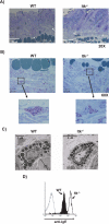
Figure 2. Normal tissue mast cells but increased expression of FcεR in Itk−/− mast cells
(A) Sections of skin from WT and Itk−/− mice analyzed by Toluidine Blue staining. Arrows indicate mast cells, with the epidermis at the top of the figure (20X). (B) Sections of skin from WT and Itk−/− mice analyzed by Toluidine Blue staining (60X). (Inset: amplified view of Toluidine Blue stained mast cells) (C) EM analysis of skin mast cells from WT and Itk−/− mice (representative images from at least 3 mice each). (D) FcεR expression on peritoneal mast cells from 6−8 week old WT (shaded lines) or Itk−/− mice (open lines) (repeated at least 4X).
Figure 3
Figure 3. Itk−/− mice have higher class switch to IgE and increased occupancy of the FcεR on their mast cells
(A) Total serum IgE levels from WT and Itk−/− analyzed by ELISA (n=6, *p<0.05) (B) Splenic B cells from 6−8 week old WT (left) or Itk−/− mice (right) were analyzed for IgE/IgM expression by gating on B220+ cells (repeated at least 3X). (C) IgE binding to FcεRI on peritoneal mast cells from 6−8 week old WT mice (left) and Itk−/− mice (right) before (control, shaded lines) and after incubation with IgE antibody (IgE, open lines) (repeated 2X).
Figure 4
Figure 4. The defect in histamine secretion in Itk−/− mice is rescued by transfer of Itk−/− BMMCs into W/Wv mice
. (A) BMMCs were expanded from bone marrow of WT and Itk−/− animals as described in materials and methods section. Surface expression of the high affinity FcεR was then determined by IgE binding and flow cytometry. Open lines, control staining in the absence of addition of IgE, filled lines, staining in the presence of IgE. (B) Surface expression of the high affinity FcεR determined by flow cytometric analysis using anti-FcεRI antibodies. (C) WT and Itk−/− BMMCs were transferred into W/Wv mice and allowed to reconstitute for 9 weeks. Histamine release was analyzed as described in Figure 1 (n=3, *p<0.05).
Figure 5
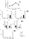
Figure 5. Normal degranulation response but enhanced cytokine secretion by Itk−/− BMMCs in response to FcεRI triggering
(A) WT or Itk−/− BMMCs were left untreated (control) or coated with anti-DNP IgE (IgE), followed by stimulation with increasing doses DNP-HSA. Supernatants were analyzed for β-hexosaminidase as a measure of degranulation. Results are expressed as a percentage of total cellular β-hexosaminidase (n=3, *p<0.05 vs. WT). (B & C) WT or Itk−/− BMMCs were left untreated (control), or coated with anti-DNP IgE (IgE), followed by stimulation with 100 ng/ml DNP-HSA (B & C) or anti-IgE (C). Supernatants were analyzed by ELISA for IL-13 (n=3, *p<0.05 vs. WT) or TNF-α (n=3, *p<0.05 vs. WT). (D) WT or Itk−/− BMMCs were incubated overnight with anti-DNP IgE alone and supernatants analyzed for the indicated cytokines (n=3, *p<0.05 vs. WT). (E) FcεRI expression on BMMCs after overnight incubation with IgE.
Figure 6
Figure 6. Calcium responses to FcεRI triggering in Itk−/− BMMCs
(A) WT or Itk−/− BMMCs were loaded with anti-DNP IgE overnight, loaded with Fura-2, then stimulated with the indicated concentration of DNP-HSA and calcium responses analyzed. Data expressed as fold increase compared to baseline signals. (B) Peak fold increase in calcium response of 2−4 experiments were averaged and plotted vs. concentration of antigenic stimulation (A & B are representative of at least 15 experiments). (C) WT or Itk−/− BMMCs were coated with anti-DNP IgE overnight, loaded with Fura-2, then stimulated with 100 ng/ml DNP-HSA in the absence of extracellular calcium at 50 sec, 2 mM calcium was added at 100 sec, Ionomycin was added as positive control at 400 sec.
Figure 7
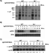
Figure 7. FcεRI mediated signaling in Itk−/− BMMCs
WT or Itk−/− BMMCs were loaded with anti-DNP IgE overnight, then stimulated with 100 ng/ml DNP-HSA for the indicated time periods. Cells were then lysed, and total cell lysates separated by SDS-PAGE, transferred to PVDF membranes and probed with anti-phosphotyrosine antibodies (A), anti-phospho-ERK (B), or anti-phospho-p38 (C)(experiments repeated at least 3X).
Figure 8
Figure 8. Increased NFAT expression and activation in Itk−/− BMMCs
(A) (Top) mRNA from WT or Itk−/− BMMCs were analyzed for the expression of the indicated NFAT isoforms by quantitative RT-PCR (n=3, experiment done on 2 separate batches of BMMCs, *p<0.05 vs. WT). (Bottom) Nonstimulated WT or Itk−/− BMMCs were analyzed for expression of NFAT2/NFATc1 by western blot. (B) Nonstimulated WT or Itk−/− BMMCs were analyzed for localization of NFAT1/NFATc2 by confocal microscopy. (Green=NFAT1; Red=DNA) (repeated 2X).

References

    1. Marone G, Triggiani M, de Paulis A. Mast cells and basophils: friends as well as foes in bronchial asthma? Trends Immunol. 2005;26:25–31. - PubMed
    1. Galli SJ, Kalesnikoff J, Grimbaldeston MA, Piliponsky AM, Williams CM, Tsai M. Mast cells as “tunable” effector and immunoregulatory cells: recent advances. Annu Rev Immunol. 2005;23:749–786. - PubMed
    1. Siraganian RP. Mast cell signal transduction from the high-affinity IgE receptor. Curr Opin Immunol. 2003;15:639–646. - PubMed
    1. Galli SJ, Nakae S, Tsai M. Mast cells in the development of adaptive immune responses. Nat Immunol. 2005;6:135–142. - PubMed
    1. Sayed BA, Brown MA. Mast cells as modulators of T-cell responses. Immunol Rev. 2007;217:53–64. - PubMed

Publication types

MeSH terms