. 2008 Jun 15;180(12):8222-30.
doi: 10.4049/jimmunol.180.12.8222.

Misoprostol impairs female reproductive tract innate immunity against Clostridium sordellii

Affiliations

Misoprostol impairs female reproductive tract innate immunity against Clostridium sordellii

David M Aronoff et al. J Immunol. .

Abstract

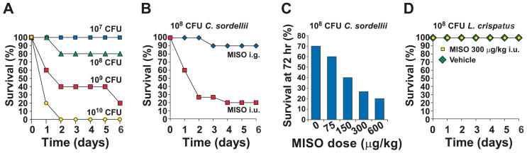
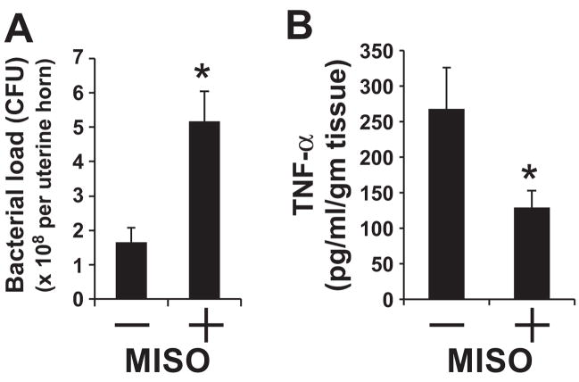
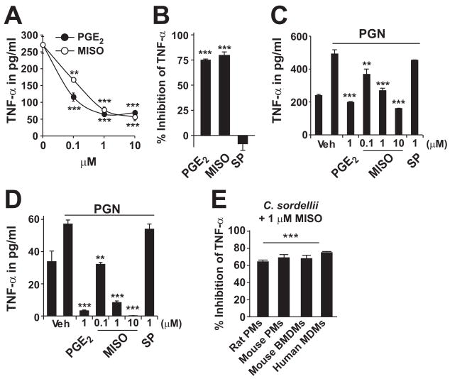
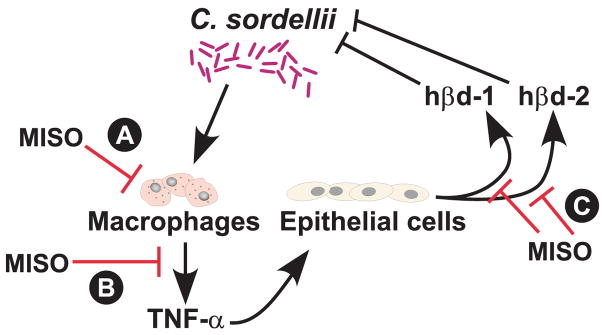
Fatal cases of acute shock complicating Clostridium sordellii endometritis following medical abortion with mifepristone (also known as RU-486) used with misoprostol were reported. The pathogenesis of this unexpected complication remains enigmatic. Misoprostol is a pharmacomimetic of PGE(2), an endogenous suppressor of innate immunity. Clinical C. sordellii infections were associated with intravaginal misoprostol administration, suggesting that high misoprostol concentrations within the uterus impair immune responses against C. sordellii. We modeled C. sordellii endometritis in rats to test this hypothesis. The intrauterine but not the intragastric delivery of misoprostol significantly worsened mortality from C. sordellii uterine infection, and impaired bacterial clearance in vivo. Misoprostol also reduced TNF-alpha production within the uterus during infection. The intrauterine injection of misoprostol did not enhance mortality from infection by the vaginal commensal bacterium Lactobacillus crispatus. In vitro, misoprostol suppressed macrophage TNF-alpha and chemokine generation following C. sordellii or peptidoglycan challenge, impaired leukocyte phagocytosis of C. sordellii, and inhibited uterine epithelial cell human beta-defensin expression. These immunosuppressive effects of misoprostol, which were not shared by mifepristone, correlated with the activation of the G(s) protein-coupled E prostanoid (EP) receptors EP2 and EP4 (macrophages) or EP4 alone (uterine epithelial cells). Our data provide a novel explanation for postabortion sepsis leading to death and also suggest that PGE(2), in which production is exaggerated within the reproductive tract during pregnancy, might be an important causal determinant in the pathogenesis of more common infections of the gravid uterus.


Conflict of interest statement

Disclosures

The authors have no financial conflict of interest.

Figures

FIGURE 1
FIGURE 1
The i.u. injection of misoprostol (MISO) worsens C. sordellii uterine infection. A, Nonpregnant rats (n = 5–7 per group) were inoculated with C. sordellii in the right uterine horn on day 0 and monitored for survival. PBS vehicle-treated rats (negative control) did not die (n = 5; data not shown). B, Rats were infected with C. sordellii (1 × 10⁸ CFU) into the right uterine horn as in A, with the administration of misoprostol (MISO, 300 μg/kg) injected into the ipsilateral uterine horn (i.u.) or stomach (i.g.). Rats per group (n = 10, i.g.) and (n = 15, i.u.) are shown. C, Rats were infected as in B with the concomitant administration of misoprostol 0 – 600 μg/kg injected i.u. into the ipsilateral uterine horn and survival was determined at 72 h after inoculation (n = 5–7 rats per group). D, Rats were infected with L. crispatus (1 × 10⁸ CFU) into the right uterine horn, with the administration of vehicle or misoprostol 300 μg/kg injected i.u. into the ipsilateral uterine horn. Rats per group (n = 5) are shown.
FIGURE 2
FIGURE 2
The i.u. injection of misoprostol (MISO) impairs bacterial clearance and TNF-α production in vivo. A, Uterine bacterial loads following infections with C. sordellii. Rats were inoculated with C. sordellii (1 × 10⁸ CFU i.u.) and misoprostol (300 μg/kg i.u.) (n = 6 rats) or vehicle-treated (n = 4 rats). Animals were sacrificed 18 h after infection and bacterial loads were determined as described in Materials and Methods. *, p < 0.05. B, Uterine TNF-α concentrations were determined 18 h after infection with C. sordellii (1 × 10⁸ CFU i.u.) in the presence of misoprostol (300 μg/kg i.u.) or vehicle (n = 6 rats per group) as described in Materials and Methods. *, p < 0.05.
FIGURE 3
FIGURE 3
Misoprostol increases cAMP in macrophages via EP2 and EP4 activation. A, Rat peritoneal macrophages were treated with PGE2 or misoprostol (MISO) at the concentrations indicated (15 min) in the presence of the phosphodiesterase inhibitor 3-isobutyl-1-methylxanthine (IBMX, 250 μM). Intracellular cAMP levels were determined by EIA. Mean values were determined in triplicate from one of three independent experiments with similar results. B, cAMP levels were determined as in A with PGE2, the EP2 agonist butaprost free acid, or the EP4 agonist Ono-AE1-329. Mean values were determined in triplicate from one of three independent experiments with similar results. **, p < 0.01 vs untreated cells.
FIGURE 4
FIGURE 4
Misoprostol impairs macrophage TNF-α production in response to LPS, PGN, or C. sordellii. A, Rat peritoneal macrophages were treated with PGE2 or misoprostol (MISO) at the concentrations indicated (60 min) before challenge with LPS (100 μg/ml) for 18 h. B, Rat peritoneal macrophages were treated with PGE2, misoprostol, or sulprostone (SP) at 10 μM concentration (60 min) before challenge with LPS (100 μg/ml) for 18 h. C, Rat peritoneal macrophages were pretreated for 1 h with PGE2, misoprostol, or sulprostone (SP) at the indicated concentrations and then exposed to PGN (10 μg/ml) for an additional 18 h. D, Rat UMs were treated as in C and supernatant TNF-α levels were determined. E, Rat peritoneal macrophages (rat PMs), mouse peritoneal macrophages (mouse PMs), mouse BMDMs, and human monocyte-derived macrophages (human MDMs) were pretreated for 1 h with misoprostol 1 μM and then exposed to heat-killed C. sordellii (at a multiplicity of infection of 100:1) for an additional 18 h. TNF-α levels were measured in cell-free supernatants and data are expressed as a percentage of inhibition of C. sordellii-treated controls in the absence of misoprostol. TNF-α levels in all experiments in cell-free supernatants were assessed by EIA. Mean data ± SEM from one of at least three independent experiments performed in triplicate. **, p < 0.01 and ***, p < 0.001 vs control.
FIGURE 5
FIGURE 5
Misoprostol inhibits macrophage phagocytosis. A, Rat peritoneal macrophages were pretreated for 5 min with increasing concentrations of misoprostol as shown and phagocytosis of FITC-labeled C. sordellii quantified by fluorometry. B, Rat peritoneal macrophages were pretreated for 5 min with increasing concentrations of misoprostol as shown, and phagocytosis of FITC-labeled E. coli opsonized with polyclonal IgG was quantified by fluorometry. Data are representative from three independent experiments performed in octuplet. Results are expressed as a percentage of relative to untreated cells. *, p = 0.05; **, p < 0.01; and ***, p < 0.001 vs control.
FIGURE 6
FIGURE 6
Misoprostol increases cAMP in UECs via EP4 receptor activation. A, ECC-1 or RL 95-2 cells were treated with PGE2 or misoprostol (MISO) at the concentrations shown for 15 min, in the presence of 3-isobutyl-1-methylxanthine (IBMX, 250 μM). Intracellular cAMP levels were measured. B, cAMP levels were determined with PGE2, the EP2 agonist butaprost free acid, or the EP4 agonist Ono-AE1-329 (all 1 μM). Mean values were determined in triplicate from one of three independent experiments with similar results.*, p < 0.05 and **, p < 0.01 vs untreated cells.
FIGURE 7
FIGURE 7
Misoprostol impairs hβd production by UECs. A, C. sordellii (104 CFU) were incubated in the presence or absence of hβd-1 or hβd-2 for 3 h under anaerobic conditions. Data shown are mean ± SEM of three independent experiments, expressed as a percentage of the growth of bacteria not exposed to defensins. B, C. sordellii were incubated as in A, in the presence or absence of 20 μg/ml hβd-2 for varying times under anaerobic conditions. Data shown are mean ± SEM of one representative experiment of two performed in triplicate. Results are expressed as a percentage of the growth of bacteria not exposed to defensins. C, Real-time PCR was used to determine the levels of mRNA for hβd-1 (relative to GAPDH) in ECC-1 and RL 95-2 cells exposed for 18 h to misoprostol (MISO, 10 μM) or vehicle. Data are mean of three determinations. D, Real-time PCR was used to determine the levels of mRNA for hβd-2 (relative to GAPDH) in TNF-α-treated (10 ng/ml) RL 95-2 cells exposed overnight to misoprostol (10 μM) or vehicle. Data are mean of three determinations. E, The hβd-2 protein levels in the cell supernatants from D were determined by EIA (n = 3). *, p < 0.05 vs untreated cells (C) or TNF-α exposed cells (D and E).
FIGURE 8
FIGURE 8
Summary of inhibitory effects of misoprostol (MISO) on female reproductive tract innate immunity. Effects of misoprostol (shown with blunt-ended red arrows) include impaired macrophage phagocytosis of C. sordellii (A), suppression of proinflammatory mediator generation in response to infection (B), and the suppression of epithelial-derived antimicrobial peptides (C).

Comment in

Similar articles

Cited by

References

    1. Fischer M, Bhatnagar J, Guarner J, Reagan S, Hacker JK, Van Meter SH, Poukens V, Whiteman DB, Iton A, Cheung M, et al. Fatal toxic shock syndrome associated with Clostridium sordellii after medical abortion. N Engl J Med. 2005;353:2352–2360. - PubMed
    1. Sinave C, Le Templier G, Blouin D, Leveille F, Deland E. Toxic shock syndrome due to Clostridium sordellii: a dramatic postpartum and post-abortion disease. Clin Infect Dis. 2002;35:1441–1443. - PubMed
    1. Cohen AL, Bhatnagar J, Reagan S, Zane SB, D’Angeli MA, Fischer M, Killgore G, Kwan-Gett TS, Blossom DB, Shieh WJ, et al. Toxic shock associated with Clostridium sordellii and Clostridium perfringens after medical and spontaneous abortion. Obstet Gynecol. 2007;110:1027–1033. - PubMed
    1. Miech RP. Pathophysiology of mifepristone-induced septic shock due to Clostridium sordellii. Ann Pharmacother. 2005;39:1483–1488. - PubMed
    1. Abramovitz M, Adam M, Boie Y, Carriere M, Denis D, Godbout C, Lamontagne S, Rochette C, Sawyer N, Tremblay NM, et al. The utilization of recombinant prostanoid receptors to determine the affinities and selectivities of prostaglandins and related analogs. Biochim Biophys Acta. 2000;1483:285–293. - PubMed

Publication types

MeSH terms