. 2008 Jun 15;180(12):8299-305.
doi: 10.4049/jimmunol.180.12.8299.

The central memory CD4+ T cell population generated during Leishmania major infection requires IL-12 to produce IFN-gamma

Affiliations

The central memory CD4+ T cell population generated during Leishmania major infection requires IL-12 to produce IFN-gamma

Nazzy Pakpour et al. J Immunol. .

Abstract

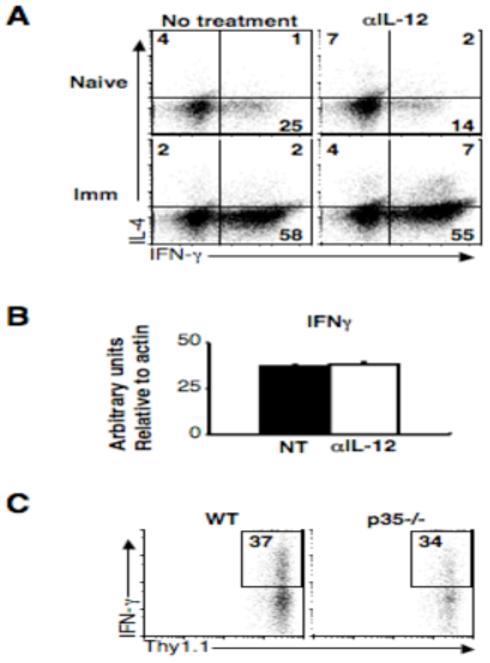
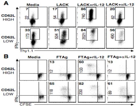
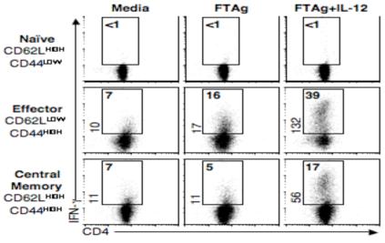
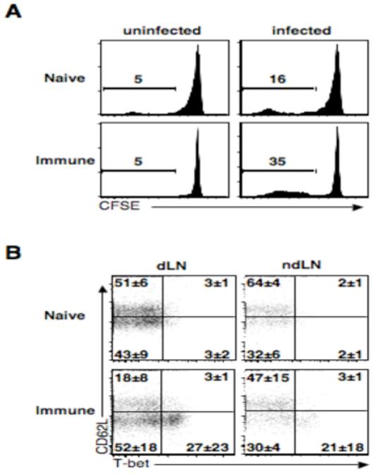
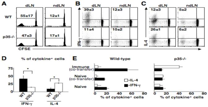
Central memory CD4(+) T cells provide a pool of lymph node-homing, Ag-experienced cells that are capable of responding rapidly after a secondary infection. We have previously described a population of central memory CD4(+) T cells in Leishmania major-infected mice that were capable of mediating immunity to a secondary infection. In this study, we show that the Leishmania-specific central memory CD4(+) T cells require IL-12 to produce IFN-gamma, demonstrating that this population needs additional signals to develop into Th1 cells. In contrast, effector cells isolated from immune mice produced IFN-gamma in vitro or in vivo in the absence of IL-12. In addition, we found that when central memory CD4(+) T cells were adoptively transferred into IL-12-deficient hosts, many of the cells became IL-4 producers. These studies indicate that the central memory CD4(+) T cell population generated during L. major infection is capable of developing into either Th1 or Th2 effectors. Thus, continued IL-12 production may be required to ensure the development of Th1 cells from this central memory T cell pool, a finding that has direct relevance to the design of vaccines dependent upon central memory CD4(+) T cells.


Figures

Figure 1
Figure 1
IL-12 at the site of infection is not required for IFN-γ production. (A) Naïve or immune C57BL/6 mice were infected and treated with 1mg αIL-12/23p40 mAb on day -1 and day 3, and on day 5 cells were isolated from infected ears. IL-4 and IFN-γ levels were determined by intracellular cytokine staining. Plots are gated on live CD4+ T cells and are representative of two experiments. Quadrant numbers are shown. (B) RNA was isolated from total ear dermis homogenate, reverse-transcribed and real-time quantitative PCR was performed with primers specific for IFN-γ. Graph shows arbitrary units relative to β-actin. (C) 20 ×106 CD4+ T cells from the spleens and lymph nodes of immune mice were transferred via the retro-orbital plexus into wild-type or IL-12p35-/- mice that were subsequently infected. After 5 days, cells were collected from infected ears and the percentage of IFN-γ+ cells is shown. Plots are gated on live Thy1.1+ donor cells and are representative of two experiments.
Figure 2
Figure 2
In vitro and in vivo generated Tcm cells require IL-12 in order to produce IFN-γ. (A) Splenocytes from B10.D2 LACK TCR transgenic ABLE-2 mice were stimulated in vitro for 3 days with LACK peptide, rIL-12, and αIL-4 mAb. Cells were then rested for five days, sorted based upon their CD62L expression and restimulated in the presence of feeder cells with LACK peptide with or without IL-12. Plots are gated on live CD4+ cells, numbers in bold represent the frequency of IFN-γ+ cells, numbers on axis represent MFI of IFN-γ+ cells. (B) CD4+ T cells from immune C57BL/6 mice were isolated and sorted into CD62Lhigh or CD62Llow groups, CFSE labeled, cultured at 1:5 ratio with splenocytes from naïve Thy disparate mice, and stimulated for four days with FTAg in the presence or absence of IL-12. Numbers represent percentage of CD4+ IFN-γ+ cells and plots are representative of two experiments.
Figure 3
Figure 3
Central memory CD4+ T cells generated by Leishmania infection require IL-12 to become IFN-γ producers. CD4+ T cells from immune C57BL/6 mice were sorted into naïve (CD62Lhigh CD44low), effector (CD62Llow CD44high), or central memory (CD62Lhigh CD44high) and cultured at a 1:5 ratio with splenocytes from naïve Thy disparate animals, and stimulated with FTAg alone or in the presence of IL-12 and αIL-4. After 4 days cells were harvested and stained for CD4 and IFN-γ. Numbers represent percentage of CD4+ IFN-γ+ cells and plots are representative of two experiments.
Figure 4
Figure 4
Central memory CD4+ T cells do not express high levels of T-bet. CD4+ T cells from immune mice were labeled with CFSE and transferred into wild-type mice that were subsequently infected and two weeks later donor cells from the draining and non-draining lymph nodes were isolated and analyzed by flow cytometry. (A) Plots are gated on live CD4+ donor T cells. Numbers represent percentage of cells that have diluted CFSE. Plots are indicative of three experiments (B) Plots are gated on proliferated live CD4+ donor T cells. Average quadrant numbers ± SD are shown.
Figure 5
Figure 5
In the absence of IL-12, the Tcm cell population becomes IL-4 producers. CD4+ T cells from immune mice (Thy1.1) were CFSE labeled and transferred into wild-type or IL-12p35-/- mice that were subsequently infected and two weeks later donor cells were isolated and analyzed by flow cytometry. (A) Numbers represent average percentage of cells that had diluted CFSE ± SD. (B) Numbers represent average percentage of proliferated IFN-γ+ ± SD. (C) Numbers represent average percentage of proliferated IL-4+ ± SD. Plots are representative of three experiments, three mice per group (D) CD4+ CD62Lhigh T cells from immune mice (Thy1.1) were sorted and transferred into wild-type or IL-12p35-/- mice that were subsequently infected, two weeks later donor cells were isolated from the dLN and analyzed by flow cytometry. Graph represents average percent of proliferated cells that are cytokine positive ± SD. *=p < 0.05 by T-test. (E) CD4+ T cells from immune (Thy1.1, CD45.2) and/or naïve C57BL/6 mice (CD45.1) were transferred into wild-type or IL-12p35-/- mice (Thy1.2, CD45.2) that were subsequently infected and two weeks later donor cells from the dLNs were isolated and analyzed by flow cytometry. Numbers represent average percent of proliferated cells that are cytokine positive ± SD. Plots are representative of two experiments.

References

    1. Scott P, Artis D, Uzonna J, Zaph C. The development of effector and memory T cells in cutaneous leishmaniasis: the implications for vaccine development. Immunol Rev. 2004;201:318–338. - PubMed
    1. Sacks D, Noben-Trauth N. The immunology of susceptibility and resistance to Leishmania major in mice. Nat Rev Immunol. 2002;2:845–858. - PubMed
    1. Sallusto F, Lenig D, Forster R, Lipp M, Lanzavecchia A. Two subsets of memory T lymphocytes with distinct homing potentials and effector functions. Nature. 1999;401:708–712. - PubMed
    1. Zaph C, Uzonna J, Beverley SM, Scott P. Central memory T cells mediate long-term immunity to Leishmania major in the absence of persistent parasites. Nat Med. 2004;10:1104–1110. - PubMed
    1. Park AY, Hondowicz B, Kopf M, Scott P. The role of IL-12 in maintaining resistance to Leishmania major. J Immunol. 2002;168:5771–5777. - PubMed

Publication types

MeSH terms