

The significance of nanoparticles in particle-induced pulmonary fibrosis
- PMID: 18523535
- PMCID: PMC2322933
The significance of nanoparticles in particle-induced pulmonary fibrosis
Abstract
Exposure to airborne nanoparticles contributes to many chronic pulmonary diseases. Nanoparticles, classified as anthropogenic and natural particles, and fibers of diameters less than 100 nm, have unrestricted access to most areas of the lung due to their size. Size relates to the deposition efficiency of the particle, with particles in the nano-range having the highest efficiencies. The deposition of nanoparticles in the lung can lead to chronic inflammation, epithelial injury, and further to pulmonary fibrosis. Cases of particle-induced pulmonary fibrosis, namely pneumoconiosis, are mostly occupationally influenced, and continue to be documented around the world. The tremendous growth of nanotechnology, however, has spurred fears of increased rates of pulmonary diseases, especially fibrosis. The severity of toxicological consequences warrants further examination of the effects of nanoparticles in humans, possible treatments and increased regulatory measures.
Keywords: Nanoparticles; asbestosis; fibrosis; nanotubes; pneumoconiosis; silicosis.
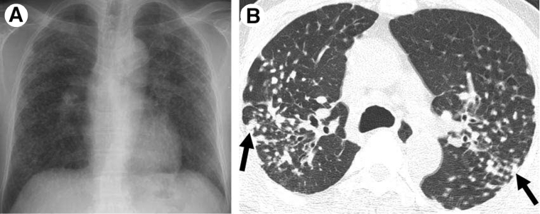
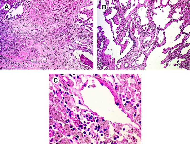
Figures
References
-
- Oberdörster G. Toxicology of Ultrafine Particles: In Vivo Studies. Philosophical Transactions of the Royal Society in London Part A. 200;358:2719–2740.
-
- Lkhasuren O, Takahashi K, Dash-Onolt L. Occupational Lung Diseases and the Mining Industry in Mongolia. International Journal of Environmental Occupational Health. 2007;13:195–201. - PubMed
-
- Wang XR, Christiani DC. Occupational Lung Disease in China. International Journal of Occupational and Environmental Health. 2003;9:320–325. - PubMed
-
- Public Health Agency of Canada. Life and Breath: Respiratory Disease in Canada. 2007. pp. 32–33.
-
- Attfield MD, Wood JM, Antao VC, Pinheiro GA. Changing Patterns of Pneumoconiosis Mortality – United States, 1968–2000. Morbidity and Mortality Weekly Report. 2004;292:795–796. - PubMed
LinkOut - more resources
Full Text Sources