. 2008 Jun 4;3(6):e2327.
doi: 10.1371/journal.pone.0002327.

The intermediate filament network in cultured human keratinocytes is remarkably extensible and resilient

Affiliations

The intermediate filament network in cultured human keratinocytes is remarkably extensible and resilient

Douglas Fudge et al. PLoS One. .

Abstract

The prevailing model of the mechanical function of intermediate filaments in cells assumes that these 10 nm diameter filaments make up networks that behave as entropic gels, with individual intermediate filaments never experiencing direct loading in tension. However, recent work has shown that single intermediate filaments and bundles are remarkably extensible and elastic in vitro, and therefore well-suited to bearing tensional loads. Here we tested the hypothesis that the intermediate filament network in keratinocytes is extensible and elastic as predicted by the available in vitro data. To do this, we monitored the morphology of fluorescently-tagged intermediate filament networks in cultured human keratinocytes as they were subjected to uniaxial cell strains as high as 133%. We found that keratinocytes not only survived these high strains, but their intermediate filament networks sustained only minor damage at cell strains as high as 100%. Electron microscopy of stretched cells suggests that intermediate filaments are straightened at high cell strains, and therefore likely to be loaded in tension. Furthermore, the buckling behavior of intermediate filament bundles in cells after stretching is consistent with the emerging view that intermediate filaments are far less stiff than the two other major cytoskeletal components F-actin and microtubules. These insights into the mechanical behavior of keratinocytes and the cytokeratin network provide important baseline information for current attempts to understand the biophysical basis of genetic diseases caused by mutations in intermediate filament genes.


Conflict of interest statement

Competing Interests: The authors have declared that no competing interests exist.

Figures

Figure 1
Figure 1. Phase contrast images of NEB-1 keratinocytes undergoing incremental uniaxial strain.
Average uniaxial strain is reported in the lower left corner of each panel. Some loss of adhesion occurs at such high strains, which is why cell strain is slightly negative when the rubber substrate is returned to its relaxed state (M). Scale bar = 50 µm.
Figure 2
Figure 2. Viability tests using the vital inclusion dye fluorescein diacetate (green) and the vital exclusion dye DAPI (blue) demonstrate that most keratinocytes survived even the most extreme uniaxial stretch treatments used in this study.
The cells shown in (A) were extended to an average maximum cell strain of 133% and then returned to the relaxed state for viability staining. Scale bar = 50 µm. (B) Average cell viability and necrosis (±SE) measured as a function of maximum cell strain reached.
Figure 3
Figure 3. Fluorescent images of NEB-1 K14-GFP keratinocytes undergoing incremental uniaxial strain.
Average uniaxial strain is reported in the lower left corner of each panel. These images demonstrate that the cytokeratin network is remarkably robust and can withstand dramatic uniaxial strains without rupturing. In some cases, keratin networks in adjacent cells separated (arrows in panel G), but this corresponded to loss of intercellular adhesion rather than a mechanical failure of the keratin network. The only visible sign of potential damage to the keratin network manifested as keratin bundles that took on a tortuous or wavy appearance after the cells were relaxed back to their resting length (arrowheads). Scale bar = 50 µm.
Figure 4
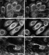
Figure 4. High magnification fluorescent images of NEB-1 K14-GFP cells before (A, C, and E) and after (B, D, and F) the cells were strained to an average maximal strain of 126%.
The main effects of this extreme uniaxial strain were increased bundling of the keratin filaments, and an increase in bundle tortuosity. Direction of stretch is indicated by the arrow in panel (A). Scale bars are 20 µm (A) and 25 µm (C) and 25 µm (E).
Figure 5
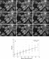
Figure 5. To measure the strain level that leads to keratin bundles adopting a wavy appearance, we loaded NEB-1 K14- GFP keratinocytes to increasingly greater maximal strains and took fluorescent images of the keratin network after the rubber substrate was returned to the unstrained state.
Average uniaxial cell strain reached is reported in the lower left corner each panel. Signs of network damage (arrowheads) appeared after cell strains as low as 50%, but became more widespread and obvious after cells were loaded to strains greater than 83%. Direction of stretch is indicated by the arrow in panel (A). Scale bar = 25 µm. (J) The tortuosity of bundles oriented parallel to the stretch axis increased significantly with the magntidue of uniaxial stretch to which the cells were subjected.
Figure 6
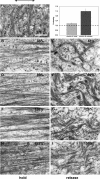
Figure 6. TEM images of keratin intermediate filaments from keratinocytes that experienced a variety of mechanical treatments before being fixed and embedded.
A) Control cells that were grown on silastic membranes and embedded without receiving any stretching treatment. All other images are from cells that were either stretched and embedded in the stretched state, or stretched and then unstretched and embedded in the relaxed state. Average maximum strain value is indicated in the upper right of each panel. The direction of stretch is indicated by the arrow above panel (A). Scale bar = 400 nm. (inset) A comparison of the tortuosity of bundles from stretched cells and cells that were stretched and relaxed demonstrates that bundles are straightened by the uniaxial strains we subjected them to.

References

    1. Coulombe PA, Wong P. Cytoplasmic intermediate filaments revealed as dynamic and multipurpose scaffolds. Nature Cell Biology. 2004;6:699–706. - PubMed
    1. Fraser RD, MacRae TP, Rogers GE. Keratins: Their Composition, Structure, and Biosynthesis. In: Kugelmass IN, editor. Springfield, IL: Charles C. Thomas; 1972. p. 304.
    1. Baribault H, Price J, Miyai K, Oshima RG. Mid-gestational lethality in mice lacking keratin 8. Genes Dev. 1993;7:1191–1202. - PubMed
    1. Fuchs E, Esteves RA, Coulombe PA. Transgenic Mice Expressing a Mutant Keratin-10 Gene Reveal the Likely Genetic-Basis for Epidermolytic Hyperkeratosis. Proceedings of the National Academy of Sciences of the United States of America. 1992;89:6906–6910. - PMC - PubMed
    1. Hesse M, Franz T, Tamai Y, Taketo MM, Magin TM. Targeted deletion of keratins 18 and 19 leads to trophoblast fragility and early embryonic lethality. Embo Journal. 2000;19:5060–5070. - PMC - PubMed

Publication types

MeSH terms

Substances