. 2008 Jun 4;3(6):e2335.
doi: 10.1371/journal.pone.0002335.

Marine actinomycetes: a new source of compounds against the human malaria parasite

Affiliations

Marine actinomycetes: a new source of compounds against the human malaria parasite

Jacques Prudhomme et al. PLoS One. .

Abstract

Background: Malaria continues to be a devastating parasitic disease that causes the death of 2 million individuals annually. The increase in multi-drug resistance together with the absence of an efficient vaccine hastens the need for speedy and comprehensive antimalarial drug discovery and development. Throughout history, traditional herbal remedies or natural products have been a reliable source of antimalarial agents, e.g. quinine and artemisinin. Today, one emerging source of small molecule drug leads is the world's oceans. Included among the source of marine natural products are marine microorganisms such as the recently described actinomycete. Members of the genus Salinispora have yielded a wealth of new secondary metabolites including salinosporamide A, a molecule currently advancing through clinical trials as an anticancer agent. Because of the biological activity of metabolites being isolated from marine microorganisms, our group became interested in exploring the potential efficacy of these compounds against the malaria parasite.

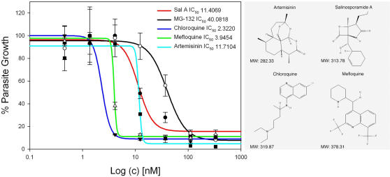
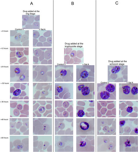
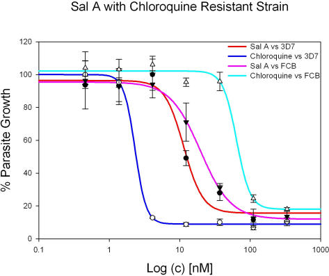
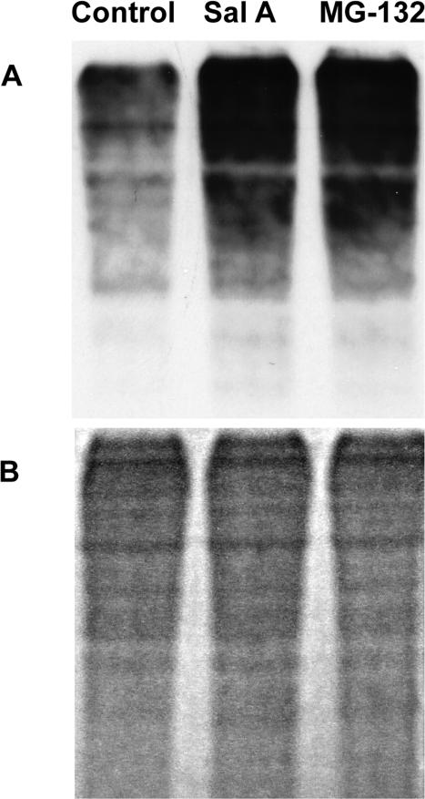
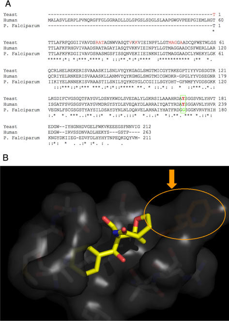
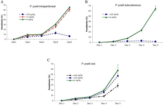
Methods: We screened 80 bacterial crude extracts for their activity against malaria growth. We established that the pure compound, salinosporamide A, produced by the marine actinomycete, Salinispora tropica, shows strong inhibitory activity against the erythrocytic stages of the parasite cycle. Biochemical experiments support the likely inhibition of the parasite 20S proteasome. Crystal structure modeling of salinosporamide A and the parasite catalytic 20S subunit further confirm this hypothesis. Ultimately we showed that salinosporamide A protected mice against deadly malaria infection when administered at an extremely low dosage.

Conclusion: These findings underline the potential of secondary metabolites, derived from marine microorganisms, to inhibit Plasmodium growth. More specifically, we highlight the effect of proteasome inhibitors such as salinosporamide A on in vitro and in vivo parasite development. Salinosporamide A (NPI-0052) now being advanced to phase I trials for the treatment of refractory multiple myeloma will need to be further explored to evaluate the safety profile for its use against malaria.


Conflict of interest statement

Competing Interests: The authors have declared that no competing interests exist.

Figures

Figure 1
Figure 1. Inhibition of P. falciparum growth by proteasome inhibitors and standard antimalarial drugs with their chemical structures.
IC50 values of parasite treated with the drug were determined using the SYBR Green assay and calculated by non linear regression using four-parameter logistic curves on SigmaPlot 10.0 software (values are mean±standard error of the mean). Each value in the curve is the average of 2 different experiments±standard deviation. Salinosporamide A inhibited the proliferation of the parasite at a low nanomolar range (IC50 = 11.4±1.9 nM) suggesting that the compound is as efficient as standard antimalarials (e.g. chloroquine (2.3±0.15 nM), mefloquine (3.9±2.7 nM) and artemisinin (11.7±8.7 nM). The inhibitor MG-132 is also a potent inhibitor of the parasites growth in vitro (IC50 of 40±4.8 nM) suggesting a strong inhibition effect of general proteasome inhibitors. Correlation coefficients (R values-factor of regression calculated by SigmaPlot 10.0) were 0.9887 for Sal A, 0.9996 for chloroquine, 0.9989 for mefloquine, 0.9900 for artemisinin and 0.9962 for MG-132.
Figure 2
Figure 2. Effect of salinosporamide A on parasite morphology.
Parasites were synchronized twice using sorbitol method. Salinosporamide A was added at the IC80 to ring (A), trophozoite (B) or schizont (C) stage. Morphological changes were observed every 6 or 12 hours by microscopic examination.
Figure 3
Figure 3. Inhibition effect of salinosporamide A on chloroquine resistant P. falciparum strain FCB.
IC50 values of parasite treated with the drug were determined using the SYBR Green assay. Each value in the curve is the average of 2 different experiments±standard deviation. Salinosporamide A inhibited the proliferation of the FCB strain suggesting that the compound is equally active against drug resistant parasites. IC50 values with Salinosporamide A were 11.4 nM±1.9 for 3D7 (R = 0.9887) and 19.6 nM±1.4 for FCB (R = 0.9990). Chloroquine IC50 values were 2.3 nM±0.15 (R = 0.9996) and 64.1 nM±4.7 (R = 0.9964) for 3D7 and FCB respectively.
Figure 4
Figure 4. Western blot analysis of parasite proteins using anti-ubiquitin antibodies.
Synchronized 3D7 parasite cultures were treated with the IC80 value of salinosporamide A (line 2) or MG-132 (Line 3). Following parasite treatment with the drug, parasite extracts were analyzed for the presence of ubiquitin-conjugates. Ubiquitinated proteins accumulate in the drugs treated when compared to the untreated cultures.
Figure 5
Figure 5
(A) Sequence alignment of the catalytic domain of the β5 subunit 20 S proteasome from yeast, human and Plasmodium obtained with the ClustalW program. (B) Crystal structure of Salinosporamide A interacting with the yeast 20S proteasome. Tyr168 is shown in orange to indicate the site of the Y168G mutation in P. falciparum.
Figure 6
Figure 6. In vivo activity of salinosporamide A against Rodent malaria parasite, P. yoellii.
Mice were treated using 3 independent delivery methods: A) Intraperitoneal B) Oral Route and C) Subcutaneous. Mice treated by oral route at 250 µg/kg (t-test, p = 0.062) or by intra peritoneal and subcutaneous methods at 130 µg/kg showed a significant decrease of parasitemia when compared with control mice (p<0.001). Parasitemia was almost cleared when used at 130 µg/kg when delivered by subcutaneous method (p<0.001). Values are means±standard deviation.

References

    1. Walker DJ, Pitsch JL, Peng MM, Robinson BL, Peters W, et al. Mechanisms of artemisinin resistance in the rodent malaria pathogen Plasmodium yoelii. Antimicrob Agents Chemother. 2000;44:344–347. - PMC - PubMed
    1. Ferrer-Rodriguez I, Perez-Rosado J, Gervais GW, Peters W, Robinson BL, et al. Plasmodium yoelii: identification and partial characterization of an MDR1 gene in an artemisinin-resistant line. J Parasitol. 2004;90:152–160. - PubMed
    1. Jambou R, Legrand E, Niang M, Khim N, Lim P, et al. Resistance of Plasmodium falciparum field isolates to in-vitro artemether and point mutations of the SERCA-type PfATPase6. Lancet. 2005;366:1960–1963. - PubMed
    1. Alker AP, Lim P, Sem R, Shah NK, Yi P, et al. Pfmdr1 and in vivo resistance to artesunate-mefloquine in falciparum malaria on the Cambodian-Thai border. Am J Trop Med Hyg. 2007;76:641–647. - PubMed
    1. Wan Y, Hur W, Cho CY, Liu Y, Adrian FJ, et al. Synthesis and target identification of hymenialdisine analogs. Chem Biol. 2004;11:247–259. - PubMed

Publication types

MeSH terms