Review
. 2008 Feb;18(1):90-100.
doi: 10.1016/j.conb.2008.05.011. Epub 2008 Jun 2.

Genetic regulation of arealization of the neocortex

Affiliations
Review

Genetic regulation of arealization of the neocortex

Dennis Dm O'Leary et al. Curr Opin Neurobiol. 2008 Feb.

Abstract

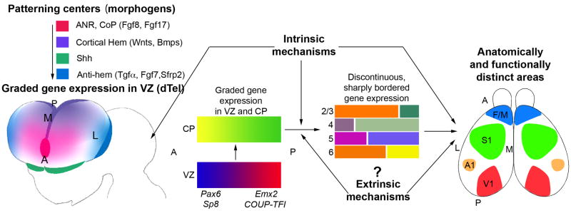
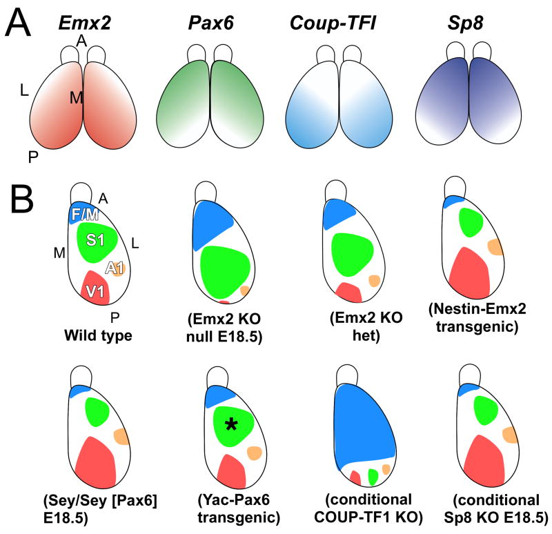
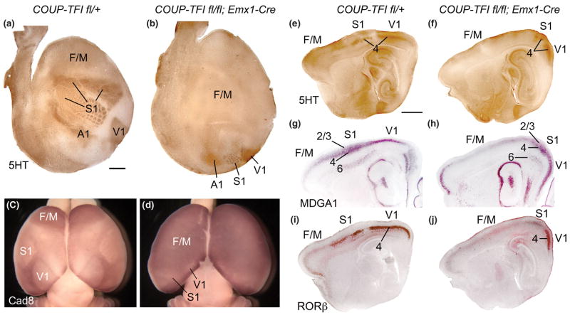
Arealization of the neocortex is controlled by a regulatory hierarchy beginning with morphogens secreted from patterning centers positioned at the perimeter of the dorsal telencephalon. These morphogens act in part to establish within cortical progenitors the differential expression of transcription factors that specify their area identity, which is inherited by their neuronal progeny, providing the genetic framework for area patterning. The two patterning centers most directly implicated in arealization are the commissural plate, which expresses fibroblast growth factors, and the cortical hem, which expresses bone morphogenetic proteins and vertebrate orthologs of Drosophila wingless, the Wnts. A third, albeit putative, patterning center is the antihem, identified by its expression of multiple signaling molecules. We describe recent findings on roles for these patterning centers in arealization. We also present the most recent evidence on functions of the four transcription factors, Emx2, COUP-TFI, Pax6, and Sp8, thus far implicated in arealization. We also describe screens for candidate target genes of these transcription factors, or other genes potentially involved in arealization. We conclude with an assessment of a forward genetics approach for identifying genes involved in determining area size based in part on quantitative trait locus mapping, and the implications for significant differences between individuals in area size on behavioral performance.


Figures

Figure 1
Figure 1. Patterning centers and graded transcription factors drive arealization of the neocortex
The initial, tangential gradients of transcription factors (TFs) in the ventricular zone (VZ) are established by signaling molecules/morphogens secreted from telencephalic patterning centers, such as Fgf8 and Fgf17 from anterior neural ridge (ANR), which later becomes the commissural plate (CoP), and Wnts and BMPs from the cortical hem. The antihem is a putative patterning center identified based on its expression of secreted signaling molecules (e.g. Tgfα, Fgf7, Sfrp2, as well as Neurogulin 1 and 3) with known patterning functions. A fourth telencephalic patterning center is defined by the expression domains of sonic hedgehog (Shh) in ventral telencephalon, but it does not have defined roles in dorsal telencephalic (dTel) patterning. The graded expression of certain TFs, such as Pax6, Emx2, COUP-TFI and Sp8, imparts positional or area identities to cortical progenitors which is imparted to their neuronal progeny that form the cortical plate (CP). The CP also initially exhibits gradients of gene expression that are gradually converted to distinct patterns with sharp borders. Coincident with this process, distinct cortical layers (2–6), and the anatomically and functionally distinct areas seen in the adult (M1, S1, A1, V1), differentiate from the CP. Genes that are differentially expressed across the cortex are often expressed in different patterns in different layers, suggesting that area-specific regulation of such genes is modulated by layer-specific properties, and questions the definition of area identity. Although the initial establishment of the graded gene expression in the embryonic CP is controlled by mechanisms intrinsic to the telencephalon, the more complex differentiation patterns established postnatally might be controlled in part by extrinsic mechanisms, for example, TCA input and the sensory activity that it relays from the periphery to the cortex. The figure is modified from [64].
Figure 2
Figure 2. Summary of graded expression of transcription factors implicated in arealization and findings in mouse mutants
(A) Graded expression in cortical progenitors of the transcription factors directly implicated in arealization, Emx2, Pax6, Coup-TFI, and Sp8, along the anterior-posterior (A–P) and lateral-medial (L–M) axes of the cortex. (B) Summary of reports of loss- or gain-of-function mutant mice of TFs that exhibit changes in area patterning. Mice with a targeted deletion of Emx2 die at birth, but late embryonic analyses suggest substantial changes in arealization as indicated in the cartoon, with a reduction in posterior areas and an expansion and posterior shift of anterior areas. Reducing Emx2 levels in the cortex of the heterozygote mutant mice (Emx2 KO het) results in posterior shifts of areas with shrinkage of V1, while overexpression of Emx2 under the control of nestin promoter (Nestin-Emx2 Transgenic) shifts areas anteriorly. Small eye mutant mice, which lack functional Pax6 protein, die at birth, but marker analyses suggest a reduction in anterior areas and an expansion and anterior shift of posterior areas. However, YAC transgenic mice of Pax6 do not show area changes other than a slight, but significant, reduction in the size of S1 (asterisk). Selective deletion of COUP-TFI in conditional knockout mice crossed with an Emx1-Cre line results in a massive expansion of frontal/motor areas and a substantial reduction of the primary sensory areas that shift posteriorly to the posterior cortical margin. Analyses of conditional knockout mice of Sp8 crossed to a BF1 (Foxg1) Cre line shows at late embryonic ages anterior shifts of gene markers, a phenotype similar to that reported for Fgf8 hypomorphic mice. The BF1-Cre line deletes Sp8 not only from cortical progenitors but also from the CoP, resulting in diminished expression of Fgf8 in the CoP. See text for details and references.
Figure 3
Figure 3. Roles and interactions between transcription factors that control arealization of the neocortex
Sp8 and Pax6 have been implicated in preferentially specifying in cortical progenitors and their progeny the identities of frontal/motor (F/M) areas, although as discussed in the text, their roles require further validation. Emx2 preferentially specifies in cortical progenitors the identities of posterior (P)/sensory (e.g. V1) areas. Coup-Tf1 represses within its more robust expression domain, the phenotypic function of any TF that may specify F/M area identities, e.g. Pax6 and Sp8 and any other TF to be identified, thereby limiting their action to anterior (A) cortical progenitors that specify F/M area identities. We also suggest based on current evidence that TFs that specify F/M area identities are dominant over the TFs that specify caudal/sensory areas and can phenotypically repress their function.
Figure 4
Figure 4. Selective deletion of COUP-TFI from cortex results in massive expansion of frontal/motor areas and posterior compression of primary sensory areas
Findings from [38] showing a prominent role for COUP-TFI in arealization. (A,B) Serotonin (5HT) immunostaining on tangential sections through layer IV of flattened cortices of P7 control (COUP-TFIfl/+) and conditional mutant (fl/fl; Emx1-Cre) cortices. Anterior is to left, and medial to the top. (A) Serotonin staining reveals primary sensory areas, including primary somatosensory (S1), visual (V1) and auditory (A1) areas, by marking area-specific TCA axon terminations. (B) In COUP-TFI fl/fl; Emx1-Cre conditional mutant brains, the primary sensory areas are much smaller than in controls and are compressed to ectopic positions at the posterior pole of the cortical hemisphere. The barrelfield of the ectopic S1 retains its characteristic patterning but is substantially reduced in size and caudally shifted, while a reduced V1 is located medial and a reduced A1 lateral to the miniature S1 barrelfield. (C,D) In situ hybridization for Cad8 on whole mounts of P7 wild-type (+/+; Emx1-Cre) and homozygous conditional mutant (COUP-TFIfl/fl; Emx1-Cre) brains uniquely marks the frontal/motor areas (F/M). The F/M areas substantially expand following selective deletion of COUP-TFI from cortex. The reduced ectopic primary sensory areas (V1, S1) can be identified by small domains of diminished cad8 expression in posterior cortex. (E–J) Serotonin (5HT) immunostaining (E,F) MDGA1 (G,H) and RORβ (I,J) in situ hybridization on serial sagittal sections of P7 control (COUP-TFIfl/+) and conditional mutant (fl/fl; Emx1-Cre) cortices. Anterior is to the left, dorsal to the top. Serotonin immunostaining reveals area-specific TCA terminations in layer 4 of S1 and V1. In conditional mutant cortex, both S1 and V1 are reduced in size and are ectopically positioned at the posterior pole of the cortical hemisphere (F). (G,H) MDGA1 selectively marks layers 4 and 6 of S1, and layer 2/3 more broadly in cortex. The S1 specific expression of MDGA1 in layers 4 and 6 confirms the reduced size and posterior shift of S1 in the COUP-TFI deficient cortex, and that these changes occur in parallel across cortical layers. (I,J) RORβ is expressed predominantly in layer 4 of the primary sensory areas (e.g. S1, V1) in wild type cortex (I). RORβ expression in the COUP-TFI deficient cortex is altered to parallel the changes in area patterning in mutant cortex (J). The majority of the cortex in the conditional mutants, including all of the neocortex anterior to the reduced, caudally-shifted primary sensory areas, exhibit serotonin staining and expression of MDGA1 and RORβ that are characteristic of wild type Frontal/Motor cortex (F/M). Scale bars: 1mm. Figure is modified from [38].

References

    1. Rakic P. Specification of cerebral cortical areas. Science. 1988;241:170–176. - PubMed
    1. O’Leary DD. Do cortical areas emerge from a protocortex? Trends Neurosci. 1989;12:400–406. - PubMed
    1. O’Leary DD, Chou SJ, Sahara S. Area patterning of the mammalian cortex. Neuron. 2007;56:252–269. - PubMed
    1. Rash BG, Grove EA. Area and layer patterning in the developing cerebral cortex. Curr Opin Neurobiol. 2006;16:25–34. - PubMed
    1. Sur M, Rubenstein JL. Patterning and plasticity of the cerebral cortex. Science. 2005;310:805–810. - PubMed

Publication types

MeSH terms

Substances