. 2008 Aug;82(16):8094-104.
doi: 10.1128/JVI.00874-08. Epub 2008 Jun 4.

Effects of lamin A/C, lamin B1, and viral US3 kinase activity on viral infectivity, virion egress, and the targeting of herpes simplex virus U(L)34-encoded protein to the inner nuclear membrane

Affiliations

Effects of lamin A/C, lamin B1, and viral US3 kinase activity on viral infectivity, virion egress, and the targeting of herpes simplex virus U(L)34-encoded protein to the inner nuclear membrane

Fan Mou et al. J Virol. 2008 Aug.

Abstract

Previous results indicated that the U(L)34 protein (pU(L)34) of herpes simplex virus 1 (HSV-1) is targeted to the nuclear membrane and is essential for nuclear egress of nucleocapsids. The normal localization of pU(L)34 and virions requires the U(S)3-encoded kinase that phosphorylates U(L)34 and lamin A/C. Moreover, pU(L)34 was shown to interact with lamin A in vitro. In the present study, glutathione S-transferase/pU(L)34 was shown to specifically pull down lamin A and lamin B1 from cellular lysates. To determine the role of these interactions on viral infectivity and pU(L)34 targeting to the inner nuclear membrane (INM), the localization of pU(L)34 was determined in LmnA(-/-) and LmnB1(-/-) mouse embryonic fibroblasts (MEFs) by indirect immunofluorescence and immunogold electron microscopy in the presence or absence of U(S)3 kinase activity. While pU(L)34 INM targeting was not affected by the absence of lamin B1 in MEFs infected with wild-type HSV as viewed by indirect immunofluorescence, it localized in densely staining scalloped-shaped distortions of the nuclear membrane in lamin B1 knockout cells infected with a U(S)3 kinase-dead virus. Lamin B1 knockout cells were relatively less permissive for viral replication than wild-type MEFs, with viral titers decreased at least 10-fold. The absence of lamin A (i) caused clustering of pU(L)34 in the nuclear rim of cells infected with wild-type virus, (ii) produced extensions of the INM bearing pU(L)34 protein in cells infected with a U(S)3 kinase-dead mutant, (iii) precluded accumulation of virions in the perinuclear space of cells infected with this mutant, and (iv) partially restored replication of this virus. The latter observation suggests that lamin A normally impedes viral infectivity and that U(S)3 kinase activity partially alleviates this impediment. On the other hand, lamin B1 is necessary for optimal viral replication, probably through its well-documented effects on many cellular pathways. Finally, neither lamin A nor B1 was absolutely required for targeting pU(L)34 to the INM, suggesting that this targeting is mediated by redundant functions or can be mediated by other proteins.


Figures
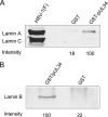
FIG. 1.
FIG. 1.
Immunoblot of lamin A/C and lamin B pulled down by GST and GST/pUL34. Full-length pUL34 fused to GST or GST alone on glutathione-Sepharose beads was reacted with HSV-1(F)-infected (A) or uninfected Hep2 cell (B) lysates, respectively. Proteins bound to GST fusion proteins were eluted and electrophoretically separated on SDS-polyacrylamide gels, transferred to nitrocellulose, and probed with either a mouse monoclonal antibody against lamin A/C (A) or goat polyclonal antibody against lamin B (B). Bound immunoglobulin was revealed by reaction with appropriate horseradish peroxidase-conjugated secondary antibodies followed by enhanced chemiluminescence. The signal intensities of protein bands were quantified and compared using Image J software, and the relative intensities of individual bands are indicated below corresponding lanes.
FIG. 2.
FIG. 2.
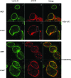
Indirect immunofluorescence localization of pUL34 in HSV-1 infected MEFs containing or lacking lamin A/C. Control MEFs or Lmna/ MEFs were infected with either wild-type HSV-1(F) (A to F) or Us3 kinase-dead mutant virus (K220A) (G to L). At 13 h postinfection, cells were fixed in paraformaldehyde and immunostained for lamin B (green) and pUL34 (red) and visualized by confocal microscopy. Optical sections for display were taken through a point between the top and bottom of cells. Insets contain magnified regions delimited by white rectangles in the same panel.
FIG. 3.
FIG. 3.
Ultrastructural localization of pUL34 in MEFs or LmnA/ MEFs infected with HSV-1(F) or US3 kinase-dead virus. (A) Wild-type MEFs were infected with HSV-1(F), and localization of pUL34 was determined by reaction with pUL34-specific IgY followed by reaction with anti-chicken immunoglobulin conjugated to 12-nm colloidal gold. Arrows indicate positions of gold beads indicative of pUL34 localization. (B and C) An experiment similar to that in panel A, except that LmnA/ MEFs were used. Colloidal gold beads are present on the INM and are indicated by arrows. (D) Immunogold localization of pUL34 in MEFs infected with the US3 kinase-dead virus. Insets show increased magnifications of the boxed areas and presence of colloidal gold beads on the INM and perinuclear virions. (E and F) Conventionally fixed images of MEFs infected with US3 kinase-dead virus showing virions within pouches of nuclear membrane. (G) Immunogold localization of pUL34 in LmnA/ cells infected with US3 kinase-dead virus. Arrows show round extensions of the nuclear membrane emanating into the cytoplasm. The inset shows a negative image of a higher magnification of the boxed area and contains an extension of the INM heavily decorated with pUL34-specific gold beads. The image is displayed as a negative to more clearly demonstrate the gold beads in white. Despite the presence of pUL34, neither virions nor nucleocapsids accumulated within the extensions of nuclear membrane.
FIG. 4.
FIG. 4.
One-step growth curve of HSV-1 on normal MEFs or LmnA/ MEFs. Cells were infected with either HSV-1(F) or Us3(K220A) at 5 PFU per cell. Residual infectivity was inactivated by a low-pH wash at 1 hour postinfection. At the indicated time points, the whole culture was collected, virus was released by freeze-thawing, and infectivity was titrated on Vero cells. Experiments were performed in duplicate. Mean values are plotted, and standard deviations are represented by error bars.
FIG. 5.
FIG. 5.
Confocal indirect immunofluorescence determination of the distribution of pUL34 in HSV-1 infected MEFs containing or lacking lamin B1. Wild-type MEFs (A and C) or LmnB1/ MEFs (B and D) were infected with HSV-1(F) or Us3(K220A) virus, fixed at 13 h postinfection, and immunostained for pUL34 as described in the legend to Fig. 2. The green channel and transmitted light from optical sections were taken near the middle of infected cells and digitally superimposed for illustrative purposes. The unaltered image was then converted to grayscale in Adobe Photoshop. Bar, 10 μM (upper panels) or 15 μM (lower panels).
FIG. 6.
FIG. 6.
Localization of pUL34 in LmnB/ cells infected with HSV-1(F) or Us3(K220A) viruses. (A) Low magnification of an HSV-1(F)-infected cell. (B) Section of nuclear membrane of cell infected with HSV-1(F). Positions of gold beads indicative of pUL34 localization are indicated by arrows. (C) Low magnification of lamin B1 knockout MEF cell infected with US3 kinase-dead virus. (D and E) Higher magnification of cells infected with US3 kinase-dead virus, showing several gold beads associated with scalloped dense-staining nuclear membrane and perinuclear virions. As a size standard, capsids are 125 nm in diameter.
FIG. 7.
FIG. 7.
Growth curves of HSV-1(F) or US3 kinase-dead virus in MEFs or LmnB/ MEFs. The experiment was performed similarly to that described in the legend to Fig. 4.

References

    1. Baines, J. D., and C. Duffy. 2006. Nucleocapsid assembly and envelopment of herpes simplex virus, p. 175-204. In R. M. Sandri-Goldin (ed.), Alpha herpesviruses: pathogenesis, molecular biology and infection control. Caister Scientific Press, Norfolk, United Kingdom.
    1. Bjerke, S. L., and R. J. Roller. 2006. Roles for herpes simplex virus type 1 U(L)34 and U(S)3 proteins in disrupting the nuclear lamina during herpes simplex virus type 1 egress. Virology 347261-276. - PMC - PubMed
    1. Chang, Y. E., C. Van Sant, P. W. Krug, A. E. Sears, and B. Roizman. 1997. The null mutant of the UL31 gene of herpes simplex virus 1: construction and phenotype of infected cells. J. Virol. 718307-8315. - PMC - PubMed
    1. Ellenberg, J., E. D. Siggia, J. E. Moreira, C. L. Smith, J. F. Presley, H. J. Worman, and J. Lippincott-Schwartz. 1997. Nuclear membrane dynamics and reassembly in living cells: targeting of an inner nuclear membrane protein in interphase and mitosis. J. Cell Biol. 1381193-1206. - PMC - PubMed
    1. Farnsworth, A., T. W. Wisner, M. Webb, R. Roller, G. Cohen, R. Eisenberg, and D. C. Johnson. 2007. Herpes simplex virus glycoproteins gB and gH function in fusion between the virion envelope and the outer nuclear membrane. Proc. Natl. Acad. Sci. USA 10410187-10192. - PMC - PubMed

Publication types