. 2008 Aug;82(16):7964-76.
doi: 10.1128/JVI.00826-08. Epub 2008 Jun 4.

Interaction of hepatitis C virus nonstructural protein 5A with core protein is critical for the production of infectious virus particles

Affiliations

Interaction of hepatitis C virus nonstructural protein 5A with core protein is critical for the production of infectious virus particles

Takahiro Masaki et al. J Virol. 2008 Aug.

Abstract

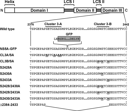
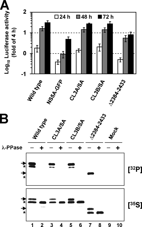
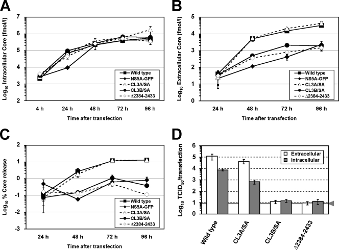
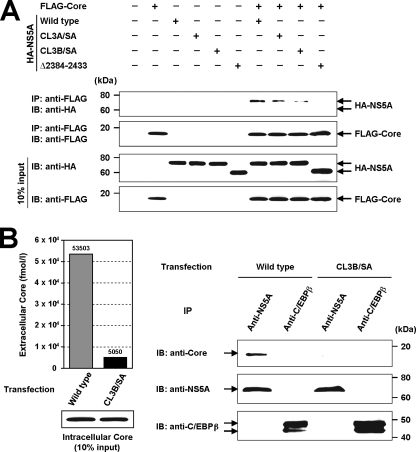
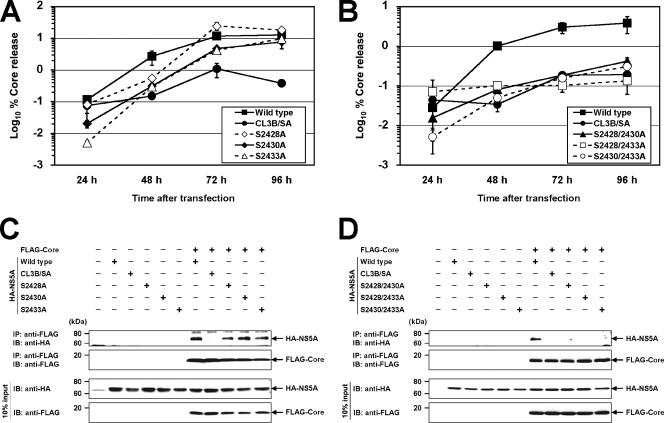
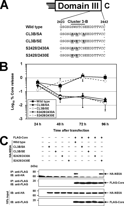
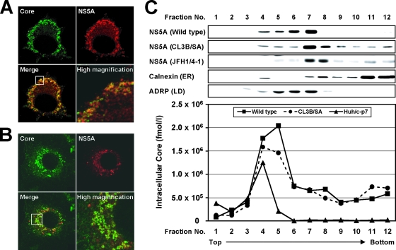
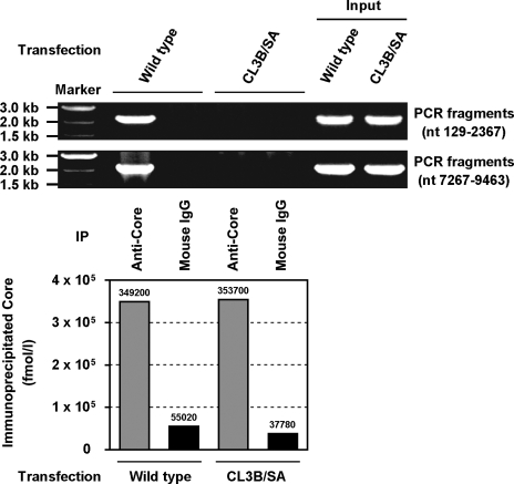
Nonstructural protein 5A (NS5A) of the hepatitis C virus (HCV) possesses multiple and diverse functions in RNA replication, interferon resistance, and viral pathogenesis. Recent studies suggest that NS5A is involved in the assembly and maturation of infectious viral particles; however, precisely how NS5A participates in virus production has not been fully elucidated. In the present study, we demonstrate that NS5A is a prerequisite for HCV particle production as a result of its interaction with the viral capsid protein (core protein). The efficiency of virus production correlated well with the levels of interaction between NS5A and the core protein. Alanine substitutions for the C-terminal serine cluster in domain III of NS5A (amino acids 2428, 2430, and 2433) impaired NS5A basal phosphorylation, leading to a marked decrease in NS5A-core interaction, disturbance of the subcellular localization of NS5A, and disruption of virion production. Replacing the same serine cluster with glutamic acid, which mimics the presence of phosphoserines, partially preserved the NS5A-core interaction and virion production, suggesting that phosphorylation of these serine residues is important for virion production. In addition, we found that the alanine substitutions in the serine cluster suppressed the association of the core protein with viral genome RNA, possibly resulting in the inhibition of nucleocapsid assembly. These results suggest that NS5A plays a key role in regulating the early phase of HCV particle formation by interacting with core protein and that its C-terminal serine cluster is a determinant of the NS5A-core interaction.


Figures

FIG. 1.
FIG. 1.
Structures of HCV constructs used in this study. Schematic diagram of the NS5A structure according to Tellinghuisen et al. (52) is shown in the top panel. The three domains are indicated by white boxes and are separated by trypsin-sensitive regions with presumably low structural complexity (low-complexity sequence [LCS]). The numbers indicate amino acid residues within the original JFH-1 polyprotein. The names listed on the left represent full-length HCV constructs, subgenomic reporter replicons, or N-terminally HA-tagged NS5A constructs used in this study. NS5A-GFP carries a GFP insertion between aa 2394 and 2395 as indicated by a shaded box. CL3A/SA and CL3B/SA carry several serine-to-alanine substitutions in the NS5A C terminus constructed as described previously (1). HCV constructs from S2428A to S2430/2433A carry single or double serine-to-alanine substitutions generated by modification of the CL3B/SA construct. The Δ2384-2433 mutant possesses an in-frame deletion in the C-terminal region of NS5A. Amino acid substitutions are marked in bold and underlined. N and C represent N terminus and C terminus, respectively.
FIG. 2.
FIG. 2.
Mutations at the C terminus of NS5A impair basal phosphorylation and have only a minor impact on RNA replication. (A) Replication of given mutants in transfected Huh-7 cells as determined by luciferase reporter assays performed at 24, 48, and 72 h posttransfection (white, gray, and black bars, respectively). Values given were normalized for transfection efficiency using the luciferase activity determined 4 h after transfection, which was set to 1. Mean values of quadruplicate measurements and the standard deviations are given. (B) Phosphorylation analysis of NS5A using the vaccinia virus T7 hybrid system. NS3-to-NS5B polyprotein fragments carrying the mutations specified above the lanes were transfected into Huh-7 cells, and proteins were radiolabeled with [32P]orthophosphate or [35S]methionine and [35S]cysteine. NS5A proteins were isolated by IP and separated by SDS-PAGE (10% polyacrylamide). Mock-transfected cells served as a negative control (lanes 9 and 10). Half of the samples were treated with λ protein phosphatase (λ-PPase) (+) whereas the other half was mock treated (−) prior to SDS-PAGE. Arrows and asterisks indicate hyperphosphorylated and basally phosphorylated forms, respectively.
FIG. 3.
FIG. 3.
Effect of mutations introduced into the NS5A C terminus on the production of infectious HCV particles. (A) Intracellular levels of core protein measured at various time points after transfection. A total of 3 × 10⁶ Huh-7 cells were transfected with 10 μg of in vitro-transcribed HCV RNAs specified in the inset and resuspended in 10 ml of culture medium, after which 2-ml aliquots were seeded into each well of a six-well culture plate. The cells were harvested at different time points between 4 h and 96 h posttransfection, and then 500 μl of cell lysate per well was prepared. After centrifugation, supernatants were processed for a core protein-specific ELISA. (B) Release of core protein from cells transfected with the HCV genomes specified in the inset. Cell culture supernatants harvested from cells given in panel A were analyzed by a core protein ELISA. (C) Efficiency of core protein release from cells transfected with the HCV genomes specified in the inset. The percent core protein release (vertical axis) indicates the percentage of released core protein in relation to total core protein (the sum of intra- and extracellular core protein) calculated for each time point. (D) Infectivity of virus particles contained in supernatants and cells after transfection with mutants specified below the graph. Culture supernatants and cells were harvested 72 h posttransfection, and extracellular (white bars) and intracellular infectivity (gray bars) levels were determined by TCID50 assay. The gray line and arrowhead represent the detection limit of the limiting dilution assay. Mean values and standard deviations for at least triplicates are shown in all panels.
FIG. 4.
FIG. 4.
aa 2428, 2430, and 2433 are essential for the interaction between NS5A and the core protein. (A) Effect of mutations at the NS5A C terminus on the interaction of NS5A with the core protein. N-terminally FLAG-tagged core protein and N-terminally HA-tagged NS5A carrying defined mutations were coexpressed in Huh-7 cells and immunoprecipitated with anti-FLAG antibody. The resulting precipitates were examined by immunoblotting using anti-HA or FLAG antibody. One-tenth of the cell lysates used in IP is shown as the 10% input. (B) Interaction between NS5A and the core protein in HCV-replicating cells. Huh-7 cells were lysed 72 h after transfection of the in vitro transcript of the HCV genome (wild type or CL3B/SA) and were immunoprecipitated with anti-NS5A antibody or anti-C/EBPβ antibody as a negative control. The resulting precipitates were examined by immunoblotting using anti-core protein, NS5A, or C/EBPβ antibody. One-tenth of cell lysates used in IP was immunoblotted with anti-core protein antibody (10% input). Cell culture supernatants harvested from transfected cells were analyzed by a core protein ELISA in parallel. IB, immunoblotting.
FIG. 5.
FIG. 5.
Determination of critical amino acids responsible for virus production and the interaction of NS5A with the core protein. (A and B) Effect of single or double serine-to-alanine substitutions on virus production. After transfection of in vitro transcripts of the HCV genomes specified in the inset into Huh-7 cells, the cells and culture supernatants were harvested at the time points given, and the amounts of the core protein were determined by core protein-specific ELISA. Percent core protein release (vertical axis) indicates the percentage of released core protein in relation to total core protein (the sum of intra- and extracellular core protein) calculated for each time point. Mean values and standard deviations for at least triplicate experiments are shown. (C and D) Effect of single or double serine-to-alanine substitutions on the interaction between NS5A and the core protein. N-terminally FLAG-tagged core protein and N-terminally HA-tagged NS5A carrying defined mutations were coexpressed in Huh-7 cells and immunoprecipitated with anti-FLAG antibody. The resulting precipitates were examined by immunoblotting using anti-HA or FLAG antibody. One-tenth of the cell lysates used in IP is shown as the 10% input. IB, immunoblotting.
FIG. 6.
FIG. 6.
Effect of glutamic acid substitutions for phosphoserines at aa 2428, 2430, and 2433 on virus production and the interaction of NS5A with the core protein. (A) Alanine or glutamic acid substitutions for serine residues at aa 2428, 2430, and 2433. The numbers indicate amino acid positions within the polyprotein of the JFH-1 isolate. The names shown on the left represent full-length HCV or N-terminally HA-tagged NS5A constructs used in this experiment. Amino acid substitutions are marked in bold and underlined. C represents the C terminus. (B) Effect of alanine or glutamic acid substitutions on virus production. After transfection of in vitro transcripts of the HCV genomes specified in the inset into Huh-7 cells, the cells and the culture supernatants were harvested at the time points given, and the amounts of core protein were determined by core protein-specific ELISA. Percent core protein release (vertical axis) indicates the percentage of released core protein in relation to total core protein (the sum of intra- and extracellular core protein) calculated for each time point. Mean values and standard deviations for at least triplicate experiments are shown. (C) Effect of alanine or glutamic acid substitutions on the interaction between NS5A and the core protein. N-terminally FLAG-tagged core protein and N-terminally HA-tagged NS5A carrying defined mutations were coexpressed in Huh-7 cells and immunoprecipitated with anti-FLAG antibody. The resulting precipitates were examined by immunoblotting (IB) using anti-HA or FLAG antibody. One-tenth of the cell lysates used in IP is as shown as the 10% input.
FIG. 7.
FIG. 7.
Subcellular localization of NS5A and the core protein in HCV-replicating cells. Huh-7 cells were transfected with the in vitro transcript of the HCV genome, wild type (A) or CL3B/SA (B). Seventy-two hours after transfection, the cells were fixed with 4% paraformaldehyde, permeabilized with 0.3% Triton X-100, and double stained with antibodies against the core protein (green) and NS5A (red), followed by staining with an Alexa Fluor 488- or Alexa Fluor 555-conjugated antibody. High-magnification panels are enlarged images of white squares in the merge panels. (C) HCV (wild type or CL3B/SA)-replicating cells, JFH1/4-1 cells harboring a subgenomic replicon of JFH-1, or Huh/c-p7 cells stably expressing JFH-1 structural proteins were lysed by freeze-thawing, and the cell lysates were fractionated on 5 to 25% iodixanol gradients. The distributions of NS5A, calnexin (ER marker), and ADRP (LD marker) were determined by immunoblotting, and those of the core protein were examined by core protein-specific ELISA.
FIG. 8.
FIG. 8.
IP-RT-PCR of HCV-replicating cells performed to examine the association between the core protein and the HCV genome RNA. Huh-7 cells were transfected with the in vitro transcript of the HCV genome (wild type or CL3B/SA) and lysed in 500 μl of hypotonic buffer at 72 h posttransfection. After IP with an anti-core protein antibody or mouse IgG, immunoprecipitates were eluted in 100 μl of elution buffer. RNAs in immunocomplexes were isolated by acid guanidinium thiocyanate-phenol-chloroform extraction. PCR was carried out as described in Materials and Methods with primer sets amplifying the fragments of nt 129 to 2367 and nt 7267 to 9463 of the JFH-1 genome. One-tenth (10 μl) of each eluted immunoprecipitate was used for assays of the core protein amounts to ensure IP efficiency (lower panel). RNA extracted from a small aliquot of each cell lysate used in IP-RT-PCR is shown as the input.

References

    1. Appel, N., T. Pietschmann, and R. Bartenschlager. 2005. Mutational analysis of hepatitis C virus nonstructural protein 5A: potential role of differential phosphorylation in RNA replication and identification of a genetically flexible domain. J. Virol. 793187-3194. - PMC - PubMed
    1. Appel, N., M. Zayas, S. Miller, J. Krijnse-Locker, T. Schaller, P. Friebe, S. Kallis, U. Engel, and R. Bartenschlager. 2008. Essential role of domain III of nonstructural protein 5A for hepatitis C virus infectious particle assembly. PLoS Pathog. 4e1000035. - PMC - PubMed
    1. Bürckstümmer, T., M. Kriegs, J. Lupberger, E. K. Pauli, S. Schmittel, and E. Hildt. 2006. Raf-1 kinase associates with hepatitis C virus NS5A and regulates viral replication. FEBS Lett. 580575-580. - PubMed
    1. Chang, K. S., J. Jiang, Z. Cai, and G. Luo. 2007. Human apolipoprotein E is required for infectivity and production of hepatitis C virus in cell culture. J. Virol. 8113783-13793. - PMC - PubMed
    1. Evans, M. J., C. M. Rice, and S. P. Goff. 2004. Phosphorylation of hepatitis C virus nonstructural protein 5A modulates its protein interactions and viral RNA replication. Proc. Natl. Acad. Sci. USA 10113038-13043. - PMC - PubMed

Publication types

LinkOut - more resources