. 2008 Sep;74(3):585-594.
doi: 10.1124/mol.108.048884. Epub 2008 Jun 4.

Pharmacological characterization of membrane-expressed human trace amine-associated receptor 1 (TAAR1) by a bioluminescence resonance energy transfer cAMP biosensor

Affiliations

Pharmacological characterization of membrane-expressed human trace amine-associated receptor 1 (TAAR1) by a bioluminescence resonance energy transfer cAMP biosensor

Larry S Barak et al. Mol Pharmacol. 2008 Sep.

Abstract

Trace amines are neurotransmitters whose role in regulating invertebrate physiology has been appreciated for many decades. Recent studies indicate that trace amines may also play a role in mammalian physiology by binding to a novel family of G protein-coupled receptors (GPCRs) that are found throughout the central nervous system. A major obstacle impeding the careful pharmacological characterization of trace amine associated receptors (TAARs) is their extremely poor membrane expression in model cell systems, and a molecular basis for this phenomenon has not been determined. In the present study, we show that the addition of an asparagine-linked glycosylation site to the N terminus of the human trace amine associated receptor 1 (TAAR1) is sufficient to enable its plasma membrane expression, and thus its pharmacological characterization with a novel cAMP EPAC (exchange protein directly activated by cAMP) protein based bioluminescence resonance energy transfer (BRET) biosensor. We applied this novel cAMP BRET biosensor to evaluate the activity of putative TAAR1 ligands. This study represents the first comprehensive investigation of the membrane-expressed human TAAR1 pharmacology. Our strategy to express TAARs and to identify their ligands using a cAMP BRET assay could provide a foundation for characterizing the functional role of trace amines in vivo and suggests a strategy to apply to groups of poorly expressing GPCRs that have remained difficult to investigate in model systems.


Figures

Fig. 1
Fig. 1
Expression of the TAAR1 in HEK-293 Cells. A, pictured are the two variants of the human trace amine receptor that were used throughout the study. Insertion of the nine-amino-acid proximal portion of the human β2-adrenergic receptor into the N terminus of the TAAR1 was a key to stabilizing it at the plasma membrane. B, confocal micrograph of the fluorescence distribution of GFP in HEK-293 cells transfected with 5 μg of cDNA of the GFP variants of the TAAR1 without and with (βTAAR1) the β2-adrenergic receptor insert. C, Western Blot for the HA epitope of the triple HA-tagged, β2-adrenergic receptor (amino acids 1–9) modified TAAR1 variants transiently expressed in HEK-293 cells. Note that the right image represents the same blot with a lower exposure period. D, immunostaining using mouse anti-HA antibody followed by goat anti-mouse Alexa Fluor 568 secondary antibody of fixed, nonpermeabilized HEK cells transiently expressing either the βTAAR1 (top), or the βTAAR1 conjugated to GFP (bottom).
Fig. 2
Fig. 2
Expression levels, distribution, and function of the βTAAR1 in live HEK cells. A, immunofluorescence and transmitted light images of Alexa Fluor 568 immuno-stained HA-β2AR (leftmost images), βTAAR1-GFP (middle images), and βTAAR1 (rightmost images). Images using live HEK-293 cells expressing labeled receptors were acquired on a Zeiss LSM 510 confocal microscope using a 100×/1.4 numerical aperture oil objective. B, overlay of Alexa Fluor 568, anti-HA immunostaining, and GFP imaging of the βTAAR1-GFP receptor. Images were acquired as above in live HEK cells plated and transfected in 35-mm glass-bottomed dishes (Matek). C, imaging of βTAAR1-GFP receptor in live U2OS cells transfected using calcium phosphate protocol with βTAAR1-GFP receptor as in B. Receptor can be seen as enhanced fluorescence at the edge of the cells. D, translocation of β-arrestin2-GFP was measured in cells transfected with β2AR (leftmost images) or the βTAAR1 (rightmost images). Live cells were imaged after labeling with monoclonal anti-HA antibody followed by Alexa Fluor 568 goat anti-mouse antibody (top), and treatment for 30 min at 37°C with either 20 μM isoproterenol (β2AR) or 20 to 50 μM p-tyramine for the βTAAR1. β-Arrestin2-GFP translocation is shown at the bottom (green).
Fig. 3
Fig. 3
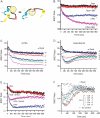
Evaluation of cAMP response in HEK-293 cells using an EPAC BRET biosensor. A, diagram of the postulated molecular rearrangement of the full-length EPAC protein with and without cAMP present. B, time course of the BRET response computed as the ratio of YFP/Rluc emissions (see Materials and Methods) in HEK-293 cells transfected with the βTAAR1 and permanently expressing the BRET biosensor (clone 8). To generate the β-PEA curve, the cells were exposed to 10 μM compound in PBS containing calcium and magnesium (see Materials and Methods), and 5 μM coelenterazine at room temperature. The lower β-PEA curve reflects the increased levels of cAMP in the presence of IBMX (0.5 mM). C, time course of the BRET sensor response to cAMP accumulation after exposure to various doses of β-PEA in clone 8 cells expressing βTAAR1 receptor. D, an analogous time course graph is generated for cAMP production secondary to isoproterenol exposure of endogenous β2-adrenergic receptors in the clone 8 HEK cells. E, the desensitizing effects of β-arrestin2 (βarrest2) cotransfection on the cAMP response of 10 μM β-PEA treated βTAAR1 results in an upward deflection of the BRET response curve. F, the Gi-coupled D2 dopamine receptor was coexpressed in HEK-293 cells expressing the EPAC BRET biosensor and endogenous β-adrenergic receptor. Increasing concentrations of dopamine preceding isoproterenol stimulation of the β-adrenergic receptors results in decreased cAMP production and deflection of the BRET response upwards toward the measured basal cAMP response profile.
Fig. 4
Fig. 4
EPAC response in HEK-293 cells with stimulation of endogenous and transfected receptors. A, clone 8 HEK-293 cells permanently expressing Rluc-EPAC-YFP and endogenous receptors only (mock) or expressing in addition transfected βTAAR1 were exposed to differing concentrations of isoproterenol or β-PEA for 5 and 15 min, respectively, at room temperature. Data are presented as the relative change ± S.E.M. from the baseline level BRET measurements, and results are the average of two (iso Mock) to four independent experiments. iso, isoproterenol. B, clone 8 cells expressing the βTAAR1 were exposed to varying concentrations of β-PEA and d-amphetamine for 15 min at room temperature. Results are presented as mean ± S.E.M. change from baseline BRET signaling and represent two independent experiments performed in duplicate. C, Dowex and Alumina column chromatography was used to measure [3H]cAMP accumulation in HEK-293 cells transfected with the βTAAR1 receptor and treated with the concentrations of compounds shown in the Figure for 15 min at room temperature. Results are the mean ± S.E.M. of two (d-amphetamine) or three (β-PEA) independent experiments performed in duplicate. D, example of TAAR1 BRET assay screening test for TAAR1 ligands. Twenty compounds were tested in this experiment at a single concentration (10 μM). Measurements were performed 10, 20, and 30 min after addition of compounds. b, vehicle baseline; compound 11, β-PEA; compound 12, l-amphetamine, compound 18, d-methamphetamine. Stippled area around the baseline represents 95% confidence interval for vehicle response. On the basis of the data presented in this report, we estimated Z-score as 0.6.

References

    1. Barak LS, Ferguson SS, Zhang J, Caron MG. A β-arrestin/green fluorescent protein biosensor for detecting G protein-coupled receptor activation. J Biol Chem. 1997;272:27497–27500. - PubMed
    1. Barak LS, Oakley RH, Laporte SA, Caron MG. Constitutive arrestin-mediated desensitization of a human vasopressin receptor mutant associated with nephrogenic diabetes insipidus. Proc Natl Acad Sci U S A. 2001;98:93–98. - PMC - PubMed
    1. Berry MD. Mammalian central nervous system trace amines. Pharmacologic amphetamines, physiologic neuromodulators. J Neurochem. 2004;90:257–271. - PubMed
    1. Borowsky B, Adham N, Jones KA, Raddatz R, Artymyshyn R, Ogozalek KL, Durkin MM, Lakhlani PP, Bonini JA, Pathirana S, et al. Trace amines: identification of a family of mammalian G protein-coupled receptors. Proc Natl Acad Sci U S A. 2001;98:8966–8971. - PMC - PubMed
    1. Boulton AA. Trace amines and mental disorders. Can J Neurol Sci. 1980;7:261–263. - PubMed

Publication types

MeSH terms

LinkOut - more resources