. 2008 Aug;100(2):1007-19.
doi: 10.1152/jn.01364.2007. Epub 2008 Jun 4.

Gustatory neural circuitry in the hamster brain stem

Affiliations

Gustatory neural circuitry in the hamster brain stem

Young K Cho et al. J Neurophysiol. 2008 Aug.

Abstract

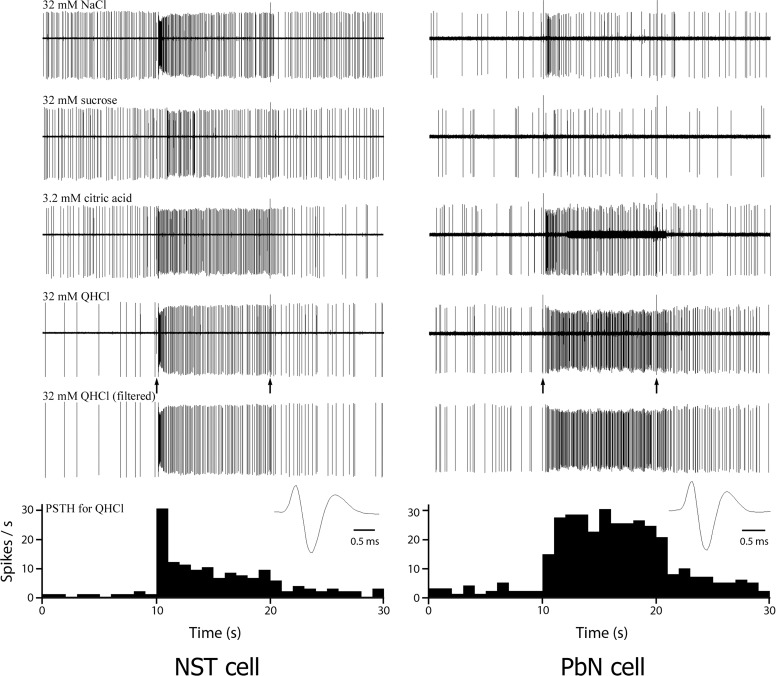
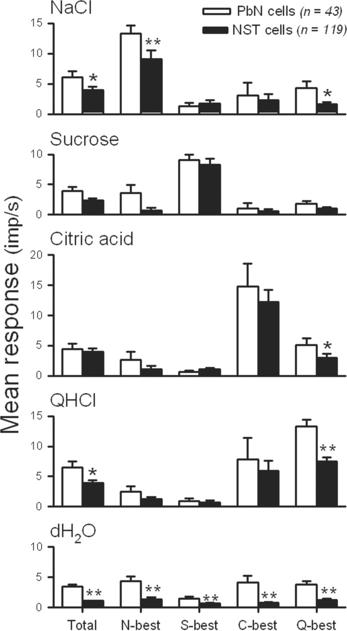
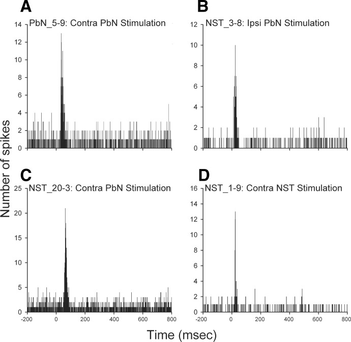
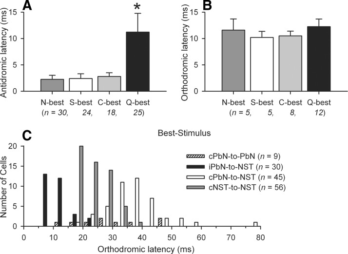
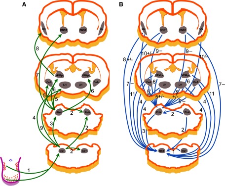
The nucleus of the solitary tract (NST) and the parabrachial nuclei (PbN) are the first and second central relays for the taste pathway, respectively. Taste neurons in the NST project to the PbN, which further transmits taste information to the rostral taste centers. Nevertheless, details of the neural connections among the brain stem gustatory nuclei are obscure. Here, we investigated these relationships in the hamster brain stem. Three electrode assemblies were used to record the activity of taste neurons extracellularly and then to electrically stimulate these same areas in the order: left PbN, right PbN, and right NST. A fourth electrode, a glass micropipette, was used to record from gustatory cells in the left NST. Results showed extensive bilateral communication between brain stem nuclei at the same level: 1) 10% of 96 NST neurons projected to the contralateral NST and 58% received synaptic input from the contralateral NST; and 2) 12% of 43 PbN neurons projected to the contralateral PbN and 21% received synaptic input from the contralateral PbN. Results also showed extensive communication between levels: 1) as expected, the majority of 119 NST neurons, 82%, projected to the ipsilateral PbN, but 85% of the 20 NST neurons tested received synaptic input from the ipsilateral PbN, as did 59% of 22 NST neurons that did not project to the PbN; and 2) although few, 3%, of 119 NST cells projected to the contralateral PbN and 38% received synaptic input from the contralateral PbN. These results demonstrated that taste neurons in the NST not only project to, but also receive descending input from the bilateral PbN and that gustatory neurons in the NST and PbN also communicate with the corresponding nucleus on the contralateral side.


Figures

FIG. 1.
FIG. 1.
Photomicrographs of stimulating and recording sites in the hamster brain stem. A: coronal section through the pons showing the position of the stimulating electrode (arrow). Iron deposit and tissue damage indicate a placement within the MPB. B: coronal section through the medulla, showing a recording site, marked with Chicago Blue dye (arrow, left NST) and a stimulating site, marked with iron deposit (arrow, right NST) within the NST. Both photomicrographs were obtained from the sections of the same animal. Abbreviations: 7, facial nucleus; DPGi, dorsal paragigantocellular nucleus; IRt, intermediate reticular nucleus; LC, locus ceruleus; Me5, mesencephalic trigeminal nucleus; Mo5, motor trigeminal nucleus; MPB, medial parabrachial nucleus; MVe, medial vestibular nucleus; NST, nucleus of the solitary tract; PCRt, parvicellular reticular nucleus; Pr, prepositus nucleus; Pr5, principal sensory trigeminal nucleus; s5, sensory root of the trigeminal nerve; scp, superior cerebellar peduncle; Sp5D, spinal trigeminal nucleus, dorsal, part; SpVe, spinal vestibular nucleus; st, solitary tract. Calibration bar = 500 μm.
FIG. 2.
FIG. 2.
Single-unit recordings from an NST (left) and a parabrachial nucleus (PbN) cell (right) in response to taste stimulation of the anterior tongue. The first 4 traces in each panel show 30-s raw recording response to NaCl, sucrose, citric acid, and QHCl stimulation, respectively. Artifacts (at 10 and 20 s) indicate opening (10 s) and closing (20 s) of the solenoids that control the delivery of the stimulus, respectively. Both neurons of the NST and PbN responded best to QHCl and the responses to QHCl of both neurons are replotted with the background activity and stimulus artifacts filtered out in each panel. The peristimulus time histograms (PSTHs) showing the impulse frequencies in response to QHCl, derived from the last recording traced in each panel (QHCl filtered), are shown at the bottom of the figure. The wave forms of the action potential of both units are shown below the last traces.
FIG. 3.
FIG. 3.
Comparison of the mean firing rate (±SE, impulses/s) of taste neurons as a function of best-stimulus category (columns) in response to the 4 taste stimuli, and distilled water (rows) in the PbN (open bars) and NST (filled bars). Total, N-best, S-best, C-best, Q-best, and dH2O correspond to the total sample, NaCl-best, sucrose-best, citric acid-best, QHCl-best, and distilled water, respectively. *P < 0.05; **P < 0.01.
FIG. 4.
FIG. 4.
Superimposed oscilloscope traces (n ≧ 3 sweeps) recorded from a taste-responsive PbN cell (A) and 3 NST neurons (B, C, and D), demonstrating fulfillment of criteria for antidromic activation from ipsilateral PbN (B), contralateral PbN (A and C), and contralateral NST (D). Responses occurred at a constant latency to the PbN (A, B, and C) and NST (D) stimuli (arrow, top), followed closely paired stimulation pulses (middle), and were canceled (▴, bottom) by collision with spontaneously generated action potentials (*). The onset latencies for antidromic activation of the units in A, B, C, and D were 3.9, 2.2, 15.2, and 18.8 ms, respectively. Scale bar = 1 ms in A and B, top traces; 2 ms in middle and bottom traces. The scale bars in units C and D represent 5 ms.
FIG. 5.
FIG. 5.
PSTHs depicting responses of a PbN taste cell (A) and 3 NST gustatory neurons (B, C, and D) following the contralateral PbN (A and C), ipsilateral PbN (B), and contralateral NST (D) stimulation, respectively. Electrical pulses were delivered at time = 0. Each PSTH was accumulated over 200 stimulus sweeps at 1/3 Hz.
FIG. 6.
FIG. 6.
A: mean antidromic latencies (±SE, ms) of the ipsilateral PbN-projecting NST taste neurons in each best-stimulus group. B: mean orthodromic latencies (±SE, ms) of the 30 NST neurons in each best-stimulus group, in response to the stimulation of the ipsilateral PbN. C: distribution of orthodromic latencies of the PbN and NST taste neurons of 4 neural connections.
FIG. 7.
FIG. 7.
Schematic diagrams illustrating the gustatory neural circuits of the rodent based on electrophysiological investigations. Note that the diagrams were based on the left (ipsilateral) side of the networks. A: projections from the recorded neuron to the stimulating site: 1: Li et al. 2008; Norgren et al. 1989; Smith et al. 1983; 2: present experiment; 3: Cho et al. 2002a; Monroe and Di Lorenzo 1995; Ogawa et al. 1984b, present experiment; 4: Cho et al. 2002b, 2003; 5: Li et al. 2005; Norgren 1974, 1976; 6: Hayama and Ogawa 1987; Mao et al. 2008; Norgren 1974, 1976; 7: Li and Cho 2006; 8: Ogawa and Nomura 1988; 9: Cho et al. 2008. B: projections from the stimulating site to the recorded neuron via synapse(s): 1: Li et al. 2008, present experiment; 2: present experiment; 3: Cho et al. 2002a, present experiment; 4: Cho et al. 2002b, 2003; Li et al. 2002; 5: Li et al. 2005; Lundy Jr and Norgren 2004a; 5′: Li et al. 2005; Lundy Jr and Norgren 2001, 2004a; 5": Lundy Jr and Norgren 2004a; 6: Mao et al. 2008; 7: Smith et al. 2005; 8: Smith and Li 2000; 9: Li and Cho 2006; 10: Di Lorenzo and Monroe 1992; Lundy Jr and Norgren 2004a; 10′: Lundy and Norgren 2004a; 11: Cho et al. 2008. The stimulation effects are indicated as follows: [−] if inhibitory responses were exclusively (9) or dominant (5", 7, 10′), [+/−] if excitatory and inhibitory responses were comparable (5, 8) or reported in different studies (5′, 10), and no symbol if the excitatory responses were exclusively (1, 2, 3) or prevailing (4, 6, 11).

References

    1. Angel A, Gratton DA. The effect of anaesthetic agents on cerebral cortical responses in the rat. Br J Pharmacol 76: 541–549, 1982 - PMC - PubMed
    1. Baird JP, Travers JB, Travers SP. Parametric analysis of gastric distension responses in the parabrachial nucleus. Am J Physiol Regul Integr Comp Physiol 281: R1568–R1580, 2001a - PubMed
    1. Baird JP, Travers SP, Travers JB. Integration of gastric distension and gustatory responses in the parabrachial nucleus. Am J Physiol Regul Integr Comp Physiol 281: R1581–R1593, 2001b - PubMed
    1. Beckman ME, Whitehead MC. Intramedullary connections of the rostral nucleus of the solitary tract in the hamster. Brain Res 557: 265–279, 1991 - PubMed
    1. Bester H, Besson JM, Bernard JF. Organization of efferent projections from the parabrachial area to the hypothalamus: a Phaseolus vulgaris-leucoagglutinin study in the rat. J Comp Neurol 383: 245–281, 1997 - PubMed

Publication types

LinkOut - more resources