Review
. 2006:117:321-39; discussion 339-40.

Placental-fetal glucose exchange and fetal glucose metabolism

Affiliations
Review

Placental-fetal glucose exchange and fetal glucose metabolism

William W Hay Jr. Trans Am Clin Climatol Assoc. 2006.

Abstract

Fetal glucose metabolism depends on additive effects of fetal plasma glucose and insulin. Glucose-stimulated insulin secretion increases over gestation, is down-regulated by constant hyperglycemia, but enhanced by pulsatile hyperglycemia. Insulin production is diminished in fetuses with intrauterine growth restriction (IUGR) by inhibition of pancreatic beta-cell replication, but not by mechanisms that regulate insulin production or secretion, while the opposite occurs with hypoglycemia alone, despite its common occurrence in IUGR. Chronic hyperglycemia down-regulates glucose tolerance and insulin sensitivity with decreased expression of skeletal muscle and hepatic Glut 1 and 4 glucose transporters, while chronic hypoglycemia up-regulates these transporters. The opposite occurs for signal transduction proteins that regulate amino acid synthesis into protein. These results demonstrate the mixed phenotype of the IUGR fetus with enhanced glucose utilization capacity, but diminished protein synthesis and growth. Such adaptations might underlie childhood and adult metabolic disorders of insulin resistance, obesity, and diabetes mellitus.


Figures

Fig. 1
Fig. 1
Uterine, fetal, and uteroplacental glucose uptake rates as functions of maternal (a, b) or fetal (c) arterial plasma glucose concentrations in pregnant sheep (9). Left Panel: Net rate of glucose transfer into the uterus from the maternal plasma versus maternal plasma glucose concentration. Vmax is the maximum rate that glucose can enter the uterus; Km is one half of Vmax and a measure of the sensitivity of uterine glucose uptake to maternal glucose concentration; Ks is the maternal glucose concentration at which Vmax is reached. Middle panel: Net rate of glucose transfer into the fetus from uteroplacenta versus maternal plasma glucose concentration; a lower fetal glucose concentration relative to any maternal glucose concentration produces a greater maternal-fetal glucose concentration gradient and a higher rate of placental to fetal glucose transfer (upper curve versus lower curve). Right panel: Net rate of uteroplacental glucose consumption versus fetal plasma glucose concentration; at any maternal glucose concentration (data for maternal glucose concentrations of 50 and 70 mg/dL are shown), uteroplacental glucose consumption is directly related to fetal glucose concentration. These data show that maternal glucose concentration determines the rate of glucose entry into the uterus (and thus the fetus and placenta), but the rate of uteroplacental glucose consumption is regulated more by the fetal than the maternal glucose concentration. Reproduced with permission from Hay, W. W. Jr.: Placental Function. In Gluckman, P., Heymann, M. A. (eds.), Scientific Basis of Pediatric and Perinatal Medicine, Second Edition, Edward Arnold Ltd., London, pps 213–227, 1996 (9); originally adapted from Hay WW Jr, Molina RD, DiGiacomo JE, Meschia G. Model of placental glucose consumption and transfer. Am J Physiol 1990;258:R569–R577 (7); Hay WW Jr, Meznarich HK. Effect of maternal glucose concentration on uteroplacental glucose consumption and transfer in pregnant sheep. Proc Soc Exp Biol Med 1988;190:63–69 (8).
Fig. 2
Fig. 2
Plasma glucose concentrations and transplacental glucose transport as functions of gestational age in pregnant sheep (10). Upper panel: Plasma glucose concentration tends to decrease in fetal sheep (-o-) over the second half of gestation relative to the maternal plasma glucose concentration (-∙-). This increases the maternal-fetal plasma glucose concentration gradient or the driving force for transplacental glucose transfer (—). Middle panel: Placental-to-fetal glucose transfer (PGT) increases about 8-fold over this same period. This increase in transport largely reflects transport capacity, shown by the increasing slope of PGT as gestational age increases. Bottom panel: At mid gestation, most of PGT is due to the maternal-fetal glucose concentration gradient whereas by term, the placental transport capacity for glucose accounts for most of PGT. Reproduced with permission from Hay, W. W. Jr. Nutrition and development of the fetus: carbohydrate and lipid metabolism. In Walker, W. A., Watkins, J. B., Duggan C. P. (eds.), Nutrition in Pediatrics (Basic Science and Clinical Applications), 3rd Edition, BC Decker Inc Publisher, Hamilton, Ontario, Canada, pp 449–470, 2003 (10). Adapted from Molina RD, Meschia G, Battaglia FC, Hay WW Jr. Maturation of placental glucose transfer capacity in the ovine pregnancy. Am J Physiol 1991;261:R697–704 (11).
Fig. 3
Fig. 3
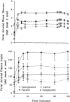
Glucose stimulated insulin secretion as a function of sustained normal, increased, or decreased plasma glucose concentrations in fetal sheep (27). Four groups of fetal sheep were studied by hyperglycemic glucose clamp technique after each group had been maintained at a unique plasma glucose concentration for 12 days: v¯ = control fetuses; ○ = fetuses that were markedly and consistently hyperglycemic (about twice normal); • = fetuses that were mildly hyperglycemic but had 3 pulses of marked hyperglycemia lasting 60 minutes each during a 24 hour period; □ = Hypoglycemic fetuses that had a plasma glucose about 50% of normal. A. fetal arterial blood glucose concentrations in 4 groups of fetal sheep during a 120 minute hyperglycemic glucose clamp. Values from 30–120 min are significantly different (P < 0.01) among all groups. B: fetal arterial plasma insulin concentrations in the same 4 groups of animals during the 120 min hyperglycemic glucose clamps. Values are means ± SEM. Reproduced with permission from Carver et al. Am J Physiol 271 (Endocrinol Metab 34):E865–E871, 1996 (27).

References

    1. Battaglia FC, Meschia G. Orlando: Academic Press; 1986. An Introduction to Fetal Physiology.
    1. Hay WW., Jr Energy and substrate requirements of the placenta and fetus.. Proc Nutr Soc; 1991. pp. 321–336. - PubMed
    1. Hay WW, Jr, Sparks JW, Quissell BJ, Battaglia FC, Meschia G. Simultaneous measurements of umbilical uptake, fetal utilization rate, and fetal turnover rate of glucose. Am J Physiol. 1981;240(6):E662–E668. - PubMed
    1. Hay WW, Jr, Sparks JW, Wilkening RB, Battaglia FC, Meschia G. Fetal glucose uptake and utilization as functions of maternal glucose concentration. Am J Physiol. 1984;246(3 Pt 1):E237–E242. - PubMed
    1. Johnson LW, Smith CH. Monosaccharide transport across microvillous membrane of human placenta. Am J Physiol. 1980;238(5):C160–C168. - PubMed

Publication types

LinkOut - more resources