

Review
doi: 10.1111/j.1600-0854.2008.00775.x.
Epub 2008 Jun 4.
Secretory outposts for the local processing of membrane cargo in neuronal dendrites
Affiliations
- PMID: 18532987
- PMCID: PMC2572994
- DOI: 10.1111/j.1600-0854.2008.00775.x
Item in Clipboard
Review
Secretory outposts for the local processing of membrane cargo in neuronal dendrites
Traffic.
2008 Sep.
Abstract
The large size and geometric complexity of neuronal dendrites necessitate specialized mechanisms to both deliver postsynaptic cargo over extended distances and regulate dendritic composition on a submicron scale. Despite the fundamental importance of membrane trafficking in dendrite growth, synapse formation and plasticity, the organelles and cellular rules governing postsynaptic trafficking are only now emerging. Here we review what is currently known about dendritic secretory organelles and their role in the development, maintenance and plasticity of postsynaptic compartments.
Figures
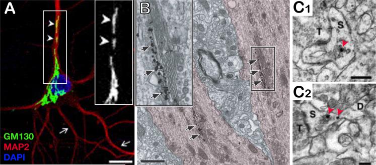
A. Immunolabeling of the somatodendritic marker MAP2 (red) and the cis-Golgi matrix protein GM130 (green) in a cultured hippocampal neuron. Scale bar, 10 μm. Inset: higher magnification of GM130 labeling illustrating Golgi outposts dispersed in the apical dendrite (arrowheads). Other dendrites lack GM130 positive Golgi outposts (arrows). B. Immunogold labeling for GM130 (arrows) demonstrating the presence of isolated Golgi stacks in the apical dendrite of a pyramidal neuron in vivo. Scale bar, 1 μm. Inset: higher magnification. A and B adapted from (24); reprinted with permission from Elsevier, copyright 2005. C. α-mannosidase II (C1) and giantin (C2) immunogold labeling (red arrowheads) in dendritic spines of CA1 pyramidal neurons in vivo. T, presynaptic terminal; S, spine; D, dendrite Scale bar, 250 nm. Adapted from (9); reprinted with permission from Elsevier, copyright 2001.
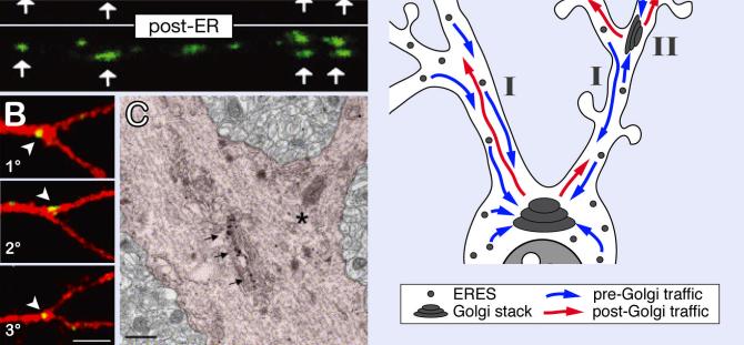
A. VSVG-ts-YFP (red) distribution in a hippocampal neuron dendrite expressing a CFP-tagged Golgi marker (GalT-CFP), before (ER), and 20 min after (post-ER) release of temperature-dependent ER exit blockade (39.5°C to 32°C switch, see text), illustrating the accumulation of post-ER cargo in dendritic Golgi outposts (arrows). Adapted from (11); reprinted with permission from the Society for Neuroscience, copyright 2003. B. VSVG-ts-GFP (green) after a 20°C TGN-exit blockade (see text) in hippocampal neurons expressing a red cell-fill, showing the presence of secretory platforms at primary (1°), secondary (2°), and tertiary (3°) dendritic branch points. C. Electron micrograph of GM130 immunogold labeling (arrows) marking a Golgi outpost located at a dendritic branch point in vivo. B and C adapted from (24); reprinted with permission from Elsevier, copyright 2005. D. Model for dual ER-to-Golgi trafficking in dendrites. While ER and ER exit sites (ERES) are distributed throughout the somatodendritic compartment, only a subset of dendrites contain Golgi outposts. Consequently, dendritic post-ER carriers are transported long distances to the somatic Golgi in dendrites lacking Golgi outposts (I). In dendrites containing Golgi outposts, post-ER carriers either bypass dendritic outposts (I) or deliver their cargo to outposts for local processing (II).

A. Three-dimensional reconstruction of serial electron micrographs showing the distribution of smooth ER (SER, dark grey) in a short segment of a CA1 hippocampal neuron dendrite in vivo. Large flat compartments (arrowheads) are linked by thin extensions (thin arrows). Note the extension of SER membranes within the head of a mature spine (crossed arrow). Adapted from (68); reprinted with permission from the Society for Neuroscience, copyright 2002. B. Electron micrograph of the spine apparatus (SA) showing the lamination of cisternae (thick arrows) between regions of high electron density (wavy arrows). Adapted from (4); reprinted with permission from the Society for Neuroscience, copyright 1997. Scale bar, 0.5 μm. C. Schematic representation of a large dendritic spine illustrating vesicles at the tip of the SA, the presence of glutamate receptors in the SA, and the presence of ribosomes (r) and polysomes (p) at the base of the spine.

A. Polarization of the somatic Golgi in pyramidal neurons (left) versus GABAergic inhibitory interneurons (right). Shown is a pseudocolored map of GM130 fluorescence intensity (Golgi-IR) as a function of radial orientation relative to the axis formed by the longest dendrite (0°, up). Insets: fluorescence fractions in each quadrant. Note that the somatic Golgi is polarized towards the apical dendrite in neurons displaying polarized dendritic trees. B. Polarization of the somatic Golgi (red) correlates with asymmetric dendrite growth to produce a single apical dendrite (1), imposing a bias to post-Golgi trafficking (blue arrows). Alteration of pre- and post-Golgi trafficking either by overexpressing dominant negative (dn) mutants of PKD or Arf1, by Sar1 RNAi knockdown, or using BFA prevents dendritic growth (1)(2). Axons are not affected under these conditions (not illustrated). Dispersion of the somatic Golgi by overexpression of GRASP65 abolishes the asymmetric growth of the apical dendrite without affecting total dendritic growth (3). Adapted from (24); reprinted with permission from Elsevier, copyright 2005.
Similar articles
-
Organelles and trafficking machinery for postsynaptic plasticity.Annu Rev Neurosci. 2006;29:325-62. doi: 10.1146/annurev.neuro.29.051605.112808. Annu Rev Neurosci. 2006. PMID: 16776589 Free PMC article. Review.
-
Secretory trafficking in neuronal dendrites.Nat Cell Biol. 2004 Jul;6(7):585-91. doi: 10.1038/ncb0704-585. Nat Cell Biol. 2004. PMID: 15232591 Review.
-
Specialization of biosynthetic membrane trafficking for neuronal form and function.Curr Opin Neurobiol. 2016 Aug;39:8-16. doi: 10.1016/j.conb.2016.03.004. Epub 2016 Mar 22. Curr Opin Neurobiol. 2016. PMID: 27010827 Review.
-
Location matters: the endoplasmic reticulum and protein trafficking in dendrites.Biol Res. 2011;44(1):17-23. doi: 10.4067/S0716-97602011000100004. Epub 2011 May 11. Biol Res. 2011. PMID: 21720677 Review.
-
Architecture and Dynamics of the Neuronal Secretory Network.Annu Rev Cell Dev Biol. 2019 Oct 6;35:543-566. doi: 10.1146/annurev-cellbio-100818-125418. Epub 2019 Jul 5. Annu Rev Cell Dev Biol. 2019. PMID: 31283381 Free PMC article. Review.
Cited by
-
Palmitoylation and depalmitoylation dynamics at a glance.J Cell Sci. 2010 Dec 1;123(Pt 23):4007-10. doi: 10.1242/jcs.059287. J Cell Sci. 2010. PMID: 21084560 Free PMC article. No abstract available.
-
Specific sets of intrinsic and extrinsic factors drive excitatory and inhibitory circuit formation.Neuroscientist. 2012 Jun;18(3):271-86. doi: 10.1177/1073858411404228. Epub 2011 Jun 7. Neuroscientist. 2012. PMID: 21652588 Free PMC article. Review.
-
The roles of protein expression in synaptic plasticity and memory consolidation.Front Mol Neurosci. 2014 Nov 12;7:86. doi: 10.3389/fnmol.2014.00086. eCollection 2014. Front Mol Neurosci. 2014. PMID: 25429258 Free PMC article. Review.
-
Translating nociceptor sensitivity: the role of axonal protein synthesis in nociceptor physiology.Eur J Neurosci. 2009 Jun;29(12):2253-63. doi: 10.1111/j.1460-9568.2009.06786.x. Epub 2009 May 29. Eur J Neurosci. 2009. PMID: 19490023 Free PMC article. Review.
-
Local zones of endoplasmic reticulum complexity confine cargo in neuronal dendrites.Cell. 2012 Jan 20;148(1-2):309-21. doi: 10.1016/j.cell.2011.11.056. Cell. 2012. PMID: 22265418 Free PMC article.
References
Publication types
MeSH terms
Substances
Grants and funding
LinkOut - more resources
Full Text Sources
