

Acute cervical fracture or congenital spinal deformity?
- PMID: 18533417
- PMCID: PMC2435025
- DOI: 10.1080/10790268.2008.11753986
Acute cervical fracture or congenital spinal deformity?
Abstract
Background/objective: There are few reports of developmental or congenital cervical spinal deformities. Such cases may be mistaken for traumatically induced fractures, and additional treatment may ensue.
Methods: A retrospective analysis was performed to identify patients with congenital cervical spine deformities. These patients were matched with a confirmed traumatic spinal fracture population with similar demographic features. Patients were analyzed for age, gender, imaging findings (plain roentgenograms including dynamic flexion and extension views, computed tomography scan, and MRI), neurologic status, and subjective complaints of pain.
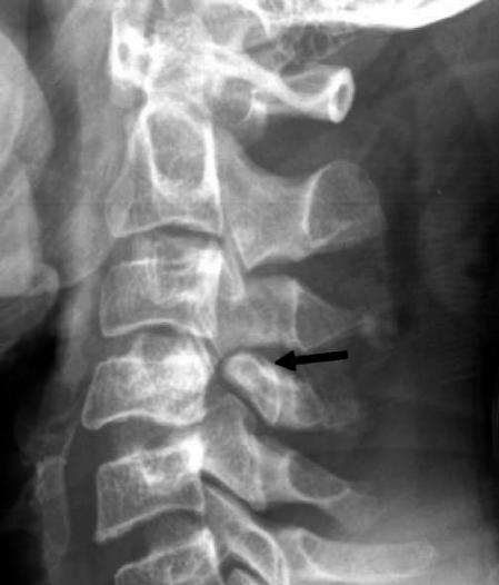
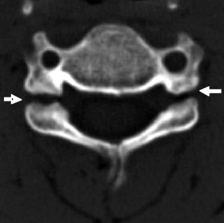
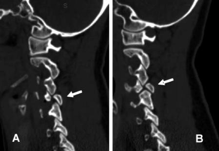
Results: Thirty-six individuals were included in the final analysis, 7 with congenital abnormalities and 29 with radiographically confirmed traumatic injuries. Patients with congenital abnormalities had significantly less soft-tissue swelling compared with the population with traumatic fractures (P < 0.001). Furthermore, those with congenital defects presented with lesser degrees of vertebral subluxation (0.29 mm vs 7.24 mm) (P < 0.0001) and without neurologic deficits (P < 0.0001).
Conclusions: Congenital abnormalities, though rare, can be mistaken for traumatic fractures of the spine. Physicians should note any evidence of soft-tissue swelling, neurologic deficits, degree of subluxation, and radiographic evidence of pedicle absence because these characteristics often provide insight into the specific etiology of the observed spinal deformity (congenital vs traumatic).
Figures
References
-
- Mantilla-Martin MT, Miller JD. Congenital absence of a pedicle in a cervical vertebra: report of two cases. Can Assoc Radiol J. 1993;44:280–284. - PubMed
-
- Bono CM, Vaccaro AR, Fehlings M, et al. Measurement techniques for lower cervical spine injuries. Spine. 2006;31((5)):603–609. - PubMed
-
- Edwards MG, Wesolowski D, Matasar K. Imaging of the absent cervical pedicle. Skeletal Radiol. 1991;20:325–328. - PubMed
-
- Gomez MA, Damie F, Besson M, Roger R, Alison D. Congenital anomaly of the cervical spine: misdiagnosis in the context of trauma (Fr) Rev Chir Orthop Reparatrice Appar Mot. 2003;89:738–741. Available at: http://srvsofcot.sofcot.com.fr/Apcort/rco/rco03/89_8/art11/art11_fs.htm. Accessed August 24, 2007. - PubMed
-
- Jones DN, Price J. Imaging of the absent cervical pedicle syndrome. Australas Radiol. 1994;38:278–281. - PubMed
MeSH terms
LinkOut - more resources
Full Text Sources
Medical