. 2008 Jun 5:9:26.
doi: 10.1186/1471-2172-9-26.

Galphas-coupled receptor signaling actively down-regulates alpha4beta1-integrin affinity: a possible mechanism for cell de-adhesion

Affiliations

Galphas-coupled receptor signaling actively down-regulates alpha4beta1-integrin affinity: a possible mechanism for cell de-adhesion

Alexandre Chigaev et al. BMC Immunol. .

Abstract

Background: Activation of integrins in response to inside-out signaling serves as a basis for leukocyte arrest on endothelium, and migration of immune cells. Integrin-dependent adhesion is controlled by the conformational state of the molecule (i.e. change in the affinity for the ligand and molecular unbending (extension)), which is regulated by seven-transmembrane Guanine nucleotide binding Protein-Coupled Receptors (GPCRs). alpha4beta1-integrin (CD49d/CD29, Very Late Antigen-4, VLA-4) is expressed on leukocytes, hematopoietic stem cells, hematopoietic cancer cells, and others. Affinity and extension of VLA-4 are both rapidly up-regulated by inside-out signaling through several Galphai-coupled GPCRs. The goal of the current report was to study the effect of Galphas-coupled GPCRs upon integrin activation.

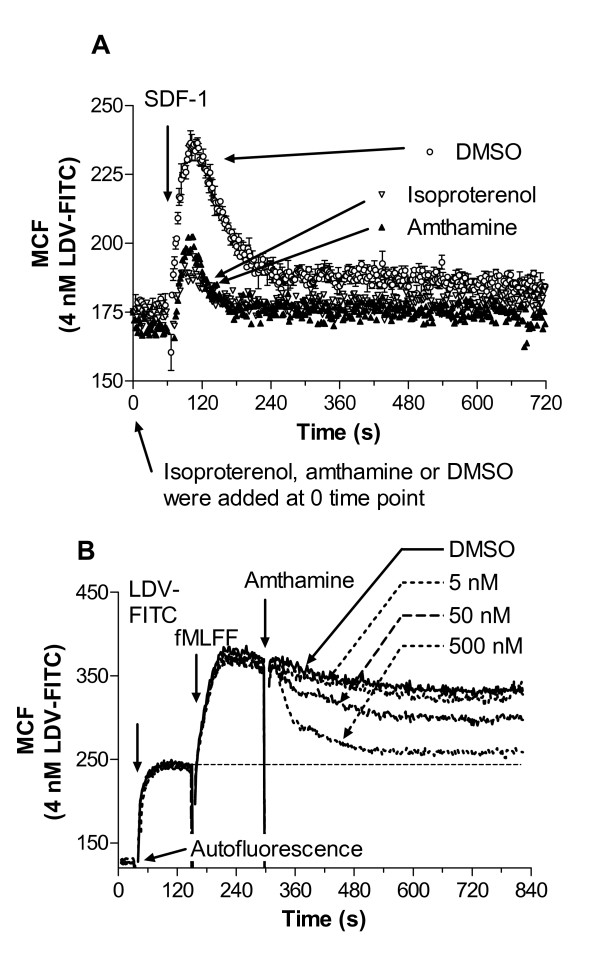
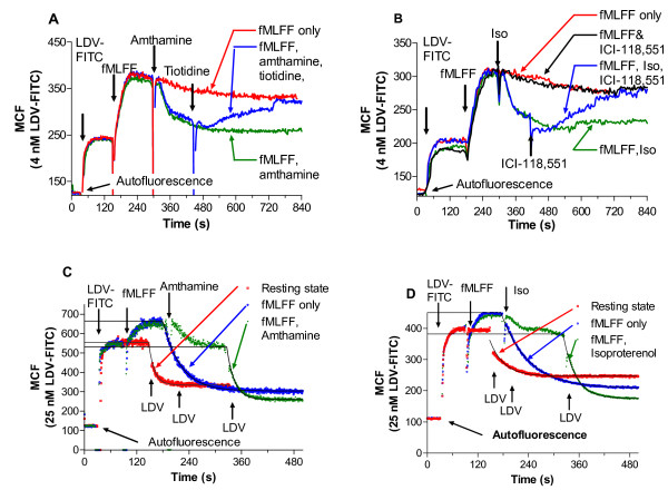
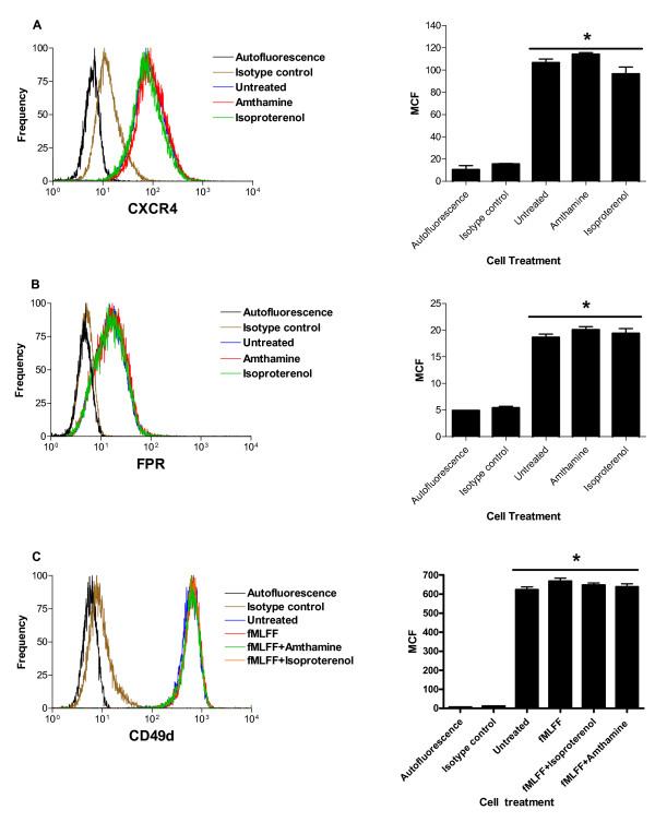
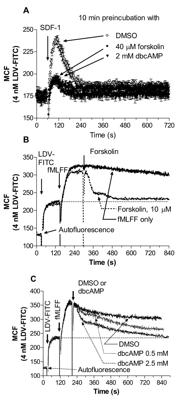
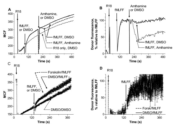
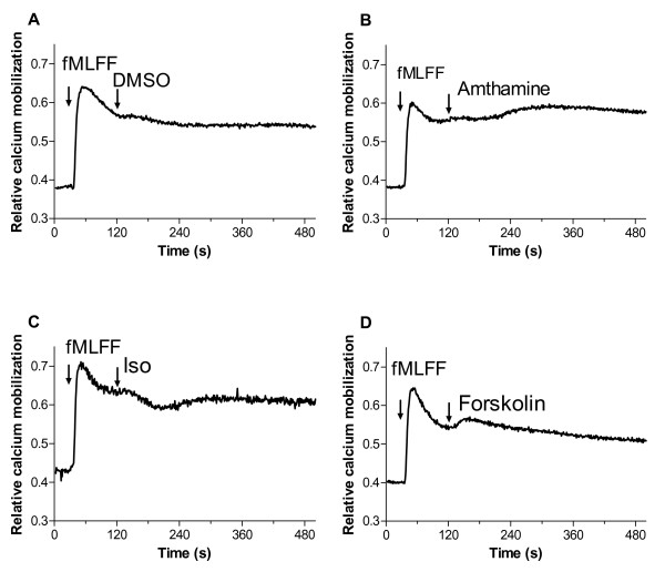
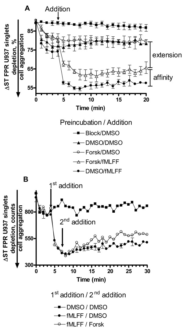
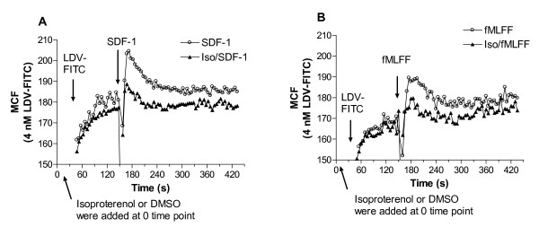
Results: Using real-time fluorescent ligand binding to assess affinity and a FRET based assay to probe alpha4beta1-integrin unbending, we show that two Galphas-coupled GPCRs (H2-histamine receptor and beta2-adrenergic receptor) as well as several cAMP agonists can rapidly down modulate the affinity of VLA-4 activated through two Galphai-coupled receptors (CXCR4 and FPR) in U937 cells and primary human peripheral blood monocytes. This down-modulation can be blocked by receptor-specific antagonists. The Galphas-induced responses were not associated with changes in the expression level of the Galphai-coupled receptors. In contrast, the molecular unbending of VLA-4 was not significantly affected by Galphas-coupled GPCR signaling. In a VLA-4/VCAM-1-specific myeloid cell adhesion system, inhibition of the VLA-4 affinity change by Galphas-coupled GPCR had a statistically significant effect upon cell aggregation.

Conclusion: We conclude that Galphas-coupled GPCRs can rapidly down modulate the affinity state of VLA-4 binding pocket through a cAMP dependent pathway. This plays an essential role in the regulation of cell adhesion. We discuss several possible implications of this described phenomenon.


Figures

Figure 1
Figure 1
Binding and dissociation of the LDV-FITC probe in response to Gαs-coupled receptor agonists. Experiments were conducted as described under "Methods". A, LDV-FITC probe binding and dissociation on U937 cells stably transfected with CXCR4 receptor plotted as mean channel fluorescence (MCF) versus time. The experiment involved sequential additions of Gαs-coupled receptor ligands (isoproterenol (100 nM) or amthamine (500 nM)), then, fluorescent LDV-FITC probe (4 nM, below saturation, added 3 min prior to addition of Gαi-coupled receptor ligands). Control cells were treated with vehicle (DMSO). Next, cells were activated with SDF-1 (25 nM, arrows). Rapid and reversible binding of the probe reflects VLA-4 affinity change [6]. Curves are means out of two independent runs calculated on a point-by-point basis. B, LDV-FITC probe binding and dissociation on U937 cells stably transfected with the non-desensitizing mutant of FPR (ΔST) [33] plotted as mean channel fluorescence (MCF) versus time. The experiment involved sequential additions of fluorescent LDV-FITC probe (4 nM), fMLFF (100 nM), and different concentrations of amthamine, or DMSO (control) (arrows). The MCF value corresponding to cell autofluorescence is indicated by the horizontal arrow. One representative experiment out of three experiments is shown. Experiments shown in the different panels were performed using different instruments, and therefore MCF values are not identical.
Figure 2
Figure 2
Binding and dissociation of the LDV-FITC probe in response to Gαs-coupled receptor agonists and antagonists. A, B, The effect of Gαs-coupled receptor antagonists. U937 cells stably transfected with the non-desensitizing mutant of FPR were sequentially treated with the LDV-FITC probe (4 nM, below saturation), fMLFF (100 nM), and A, amthamine (500 nM), tiotodine (10 μM), or B, isoproterenol (1 nM), ICI-118,551 (10 μM). B, Additional control sample was treated with the same concentration of LDV-FITC, fMLFF, and ICI-118,551 without addition of isoproterenol to show the effect of Gαs-coupled receptor antagonist without addition of the agonist. The MCF value corresponding to cell autofluorescence is indicated by the horizontal arrow. Data are plotted as MCF versus time. One representative experiment out of three experiments is shown. C,D, Kinetic analysis of binding and dissociation of LDV-FITC probe on U937 cells stably transfected with the non-desensitizing mutant of FPR. Cells were sequentially treated with the LDV-FITC probe (25 nM, near saturation), Gαi-coupled receptor ligand (fMLFF, 100 nM), Gαs-coupled receptor ligands, C, amthamine (500 nM), or D, isoproterenol (100 nM). At time points indicated by arrows, cells were treated with excess unlabeled LDV containing small molecule (2 μM), and the dissociation of the fluorescent molecule was followed. Dissociation rate constants (koff) were obtained by fitting dissociation curves to a single exponential decay equation (as described in the text). Experiments shown in the different panels were performed using different instruments, and therefore MCF values are not identical.
Figure 3
Figure 3
Effect of Gαs-coupled receptor activation upon surface expression of CXCR4 receptor, formyl peptide receptor, and VLA-4. U937 cells were treated with vehicle (untreated control), amthamine, isoproterenol, or sequentially with fMLFF, and vehicle (untreated control), amthamine or isoproterenol as described for experiments shown in Fig. 1 and 2. Next, cells were placed on ice and stained with anti-CXCR4 antibodies, A, anti-FPR antibodies, B, or CD49d antibodies, C. Histograms and bar graphs of mean channel fluorescence (MCF) ± SEM (n = 3) for unstained cell (autofluorescence), nonspecific binding (isotype control), treated with vehicle (untreated), and treated with different ligands are shown. One representative experiment out of two experiments is shown. * indicates means are not significantly different (P > 0.05) as calculated by one-way ANOVA analysis using GraphPad Prism software.
Figure 4
Figure 4
Binding and dissociation of the LDV-FITC probe in response to cAMP agonists. A, LDV-FITC probe binding and dissociation on U937 cells stably transfected with CXCR4 receptor plotted as mean channel fluorescence (MCF) versus time. Prior to SDF-1 addition, cells were preincubated with forskolin (40 μM), dibutyryl-cAMP (2 mM), or DMSO (control) for 10 min at 37°C. Next, cells were preincubated wth 4 nM LDV-FITC, and treated with SDF-1 (25 nM). Mean and standard error of mean are shown (n = 3). B, C, LDV-FITC probe binding and dissociation on U937 cells stably transfected with the non-desensitizing mutant of FPR plotted as mean channel fluorescence (MCF) versus time. After sequential additions of fluorescent LDV-FITC probe (4 nM), and fMLFF (100 nM), cells were treated with B, forskolin (10 μM), C, different concentration of dibutyryl-cAMP, or DMSO (control). The MCF value corresponding to cell autofluorescence is indicated by the horizontal arrow. One representative experiment out of three experiments is shown.
Figure 5
Figure 5
Energy transfer, on U937 cells stably transfected with the non-desensitizing mutant of FPR, between the LDV-FITC donor probe and octadecylrhodamine (R18) acceptor probe. Experiments were conducted as described under "Methods", and in [18,19]. A, Cells were preincubated at 37°C with 100 nM LDV-FITC probe to saturate high and low affinity sites. Next, LDV-FITC fluorescence was quenched after addition of octadecyl rhodamine (R18, 10 μM). Then, cells were activated by addition of fMLFF (100 nM). Data are plotted as MCF versus time for three conditions: quenched and then activated by Gαi-coupled receptor agonist (fMLFF (solid line)), quenched, then activated by Gαi-coupled receptor agonist (fMLFF) and then activated by Gαs-coupled receptor agonist (amthamine, 500 nM (dashed line)), and quenched only (R18 only, DMSO vehicle, dotted line). B, Data from panel A were re-plotted by subtracting the baseline data (R18 only) from activated cell data. The data are normalized assuming that average MCF value for FPR activated cells is equal to 100%; therefore, the Y-axis is labeled as "Donor fluorescence, % relative to fMLFF". As shown previously, activation of the cells transfected with the non-desensitizing mutant of FPR [18] led to the rapid unquenching of the FITC signal, which was interpreted as a change in the distance of closest approach between the VLA-4 ligand binding site occupied by LDV-FITC and the membrane surface (so called unbending or extension of the integrin molecule, see Fig. 1 in [18], or Fig. 5C in [19]). Curves are means out of five independent runs calculated on a point-by-point basis. C, Cells were preincubated for 5 min at 37°C with 10 μM of forskolin or DMSO (control). Next, cells were stained with 100 nM LDV-FITC probe and fluorescence was quenched after addition of octadecyl rhodamine (R18, 10 μM). Cells were activated by addition of 100 nM fMLFF, or DMSO (control). Notice the absence of the difference between forskolin treated (forskolin/fMLFF) and DMSO treated (DMSO/fMLFF) cells after addition of fMLFF. Mean and standard error of mean are shown (n = 3). D, Data from panel C were re-plotted as described for panel B.
Figure 6
Figure 6
Kinetics of intracellular Ca2+ response, detected using Indo-1, AM, in U937 cells stably transfected with the non-desensitizing mutant of FPR in response to activation by Gαi/Gαs-coupled receptor agonists. Experiments were conducted as described under "Methods". A, Control cells loaded with Indo-1 were treated with fMLFF (100 nM) and DMSO. B, Cells were sequentially treated with fMLFF (100 nM) and amthamine (500 nM); C, cells were treated with fMLFF (100 nM) and isoproterenol (100 nM); D, cells were treated with fMLFF and Forskolin (40 μM). One representative experiment out of three experiments is shown.
Figure 7
Figure 7
Changes in cell adhesion between U937 FPR (ΔST) and VCAM-1-transfected B78H1 cells at resting state and in response to receptor stimulation. A, U937 FPR (ΔST) cells were preincubated for 10 min at 37°C with forskolin (20 μM), or DMSO (vehicle). Cells were stimulated (arrow) with fMLFF (100 nM), or DMSO (vehicle). Data are plotted as % of U937 FPR (ΔST) singlets versus time. Five different experimental conditions are shown: Block/DMSO (preincubation with blocking 2 μM LDV small molecule (addition of DMSO, 1 μl), DMSO/DMSO, resting state (preincubation with DMSO/addition of DMSO), Forsk/DMSO (preincubation with forskolin/addition of DMSO), Forsk/fMLFF (preincubation with 20 μM forskolin/stimulation with 100 nM fMLFF), DMSO/fMLFF (preincubation with DMSO, stimulation with 100 nM fMLFF). The non-desensitizing FPR mutant is used to maintain VLA-4 in a state of constant high affinity. Mean and standard error of mean of three independent experiments are shown (n = 3). Bars on the right side of the panel indicate a relative contribution of affinity and extension in the overall cell adhesion. B, Effect of forskolin addition on adhesion in real-time. Cells were stimulated with fMLFF, or DMSO (1st addition). Next, forskolin (20 μM) was added. Representative experiment out of two experiments is shown.
Figure 8
Figure 8
Binding and dissociation of the LDV-FITC probe in response to Gαs-coupled receptor agonist. Experiments were conducted as described under "Methods". A, LDV-FITC probe binding and dissociation on peripheral blood monocytes plotted as mean channel fluorescence (MCF) versus time. The experiment analogous to the one shown in Fig. 1A involved sequential additions of Gαs-coupled receptor ligand (isoproterenol (100 nM)) or DMSO (control), next, fluorescent LDV-FITC probe (4 nM, below saturation, added 2 min prior to addition of Gαi-coupled receptor ligand). Cells were activated with A, SDF-1 (25 nM, arrow), or B, fMLFF (100 nM, arrow). Rapid and reversible binding of the probe reflects a transient VLA-4 affinity change. Curves are means out of four independent runs calculated on a point-by-point basis. One representative experiment out of two independent experiments for each ligand is shown. Experiments shown in the different panels were performed using different instruments, and therefore MCF values are not identical.

Similar articles

Cited by

References

    1. Hynes RO. Integrins: bidirectional, allosteric signaling machines. Cell. 2002;110:673–687. doi: 10.1016/S0092-8674(02)00971-6. - DOI - PubMed
    1. Cai W, Chen X. Anti-angiogenic cancer therapy based on integrin alphavbeta3 antagonism. Anticancer Agents Med Chem. 2006;6:407–428. doi: 10.2174/187152006778226530. - DOI - PubMed
    1. De Meyer SF, Vanhoorelbeke K, Ulrichts H, Staelens S, Feys HB, Salles I, Fontayne A, Deckmyn H. Development of monoclonal antibodies that inhibit platelet adhesion or aggregation as potential anti-thrombotic drugs. Cardiovasc Hematol Disord Drug Targets. 2006;6:191–207. doi: 10.2174/187152906778249536. - DOI - PubMed
    1. Hilden TJ, Nurmi SM, Fagerholm SC, Gahmberg CG. Interfering with leukocyte integrin activation--a novel concept in the development of anti-inflammatory drugs. Ann Med. 2006;38:503–511. doi: 10.1080/07853890600969130. - DOI - PubMed
    1. O'connor P. Natalizumab and the role of alpha 4-integrin antagonism in the treatment of multiple sclerosis. Expert Opin Biol Ther. 2007;7:123–136. doi: 10.1517/14712598.7.1.123. - DOI - PubMed

Publication types

MeSH terms

LinkOut - more resources