. 2008 Aug;212(2):348-57.
doi: 10.1016/j.expneurol.2008.04.017. Epub 2008 Apr 25.

Glutamate receptor plasticity and activity-regulated cytoskeletal associated protein regulation in the phrenic motor nucleus may mediate spontaneous recovery of the hemidiaphragm following chronic cervical spinal cord injury

Affiliations

Glutamate receptor plasticity and activity-regulated cytoskeletal associated protein regulation in the phrenic motor nucleus may mediate spontaneous recovery of the hemidiaphragm following chronic cervical spinal cord injury

Warren J Alilain et al. Exp Neurol. 2008 Aug.

Abstract

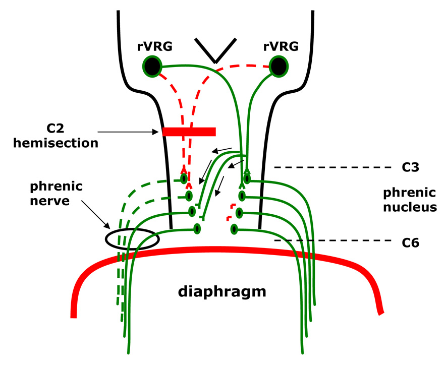
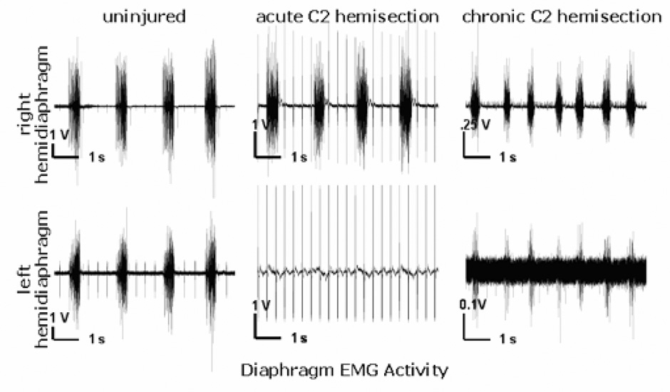
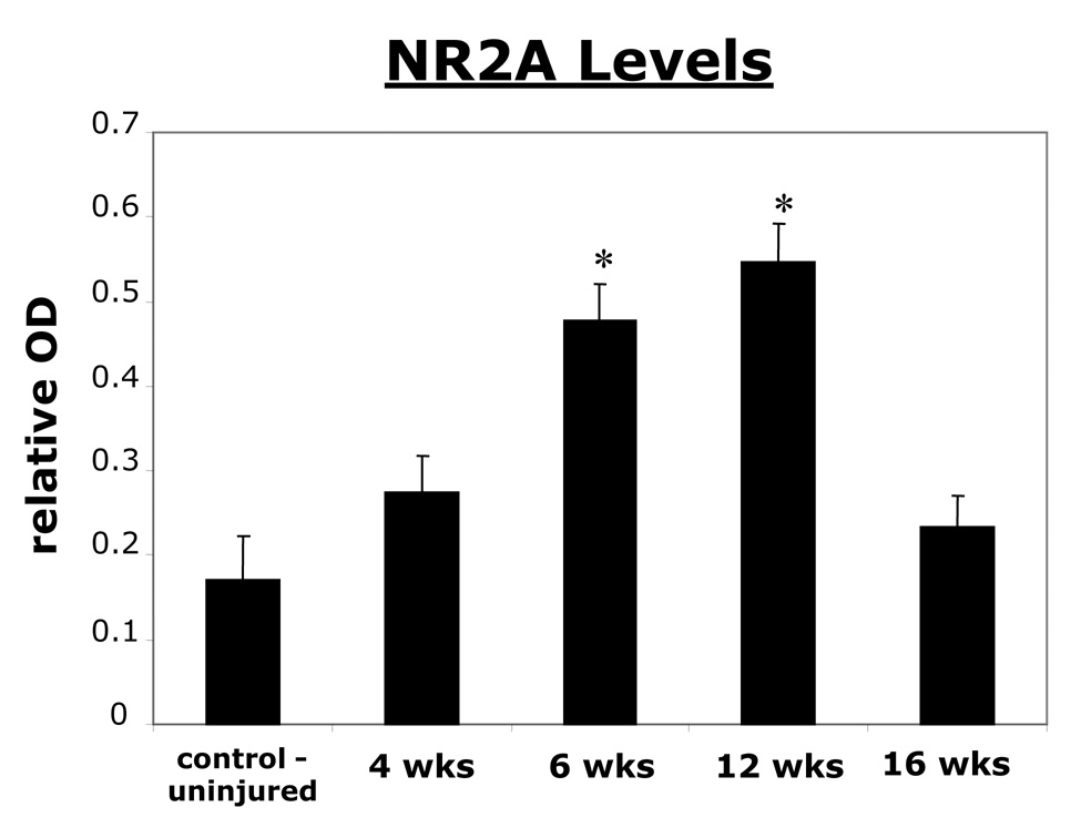
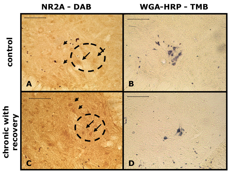
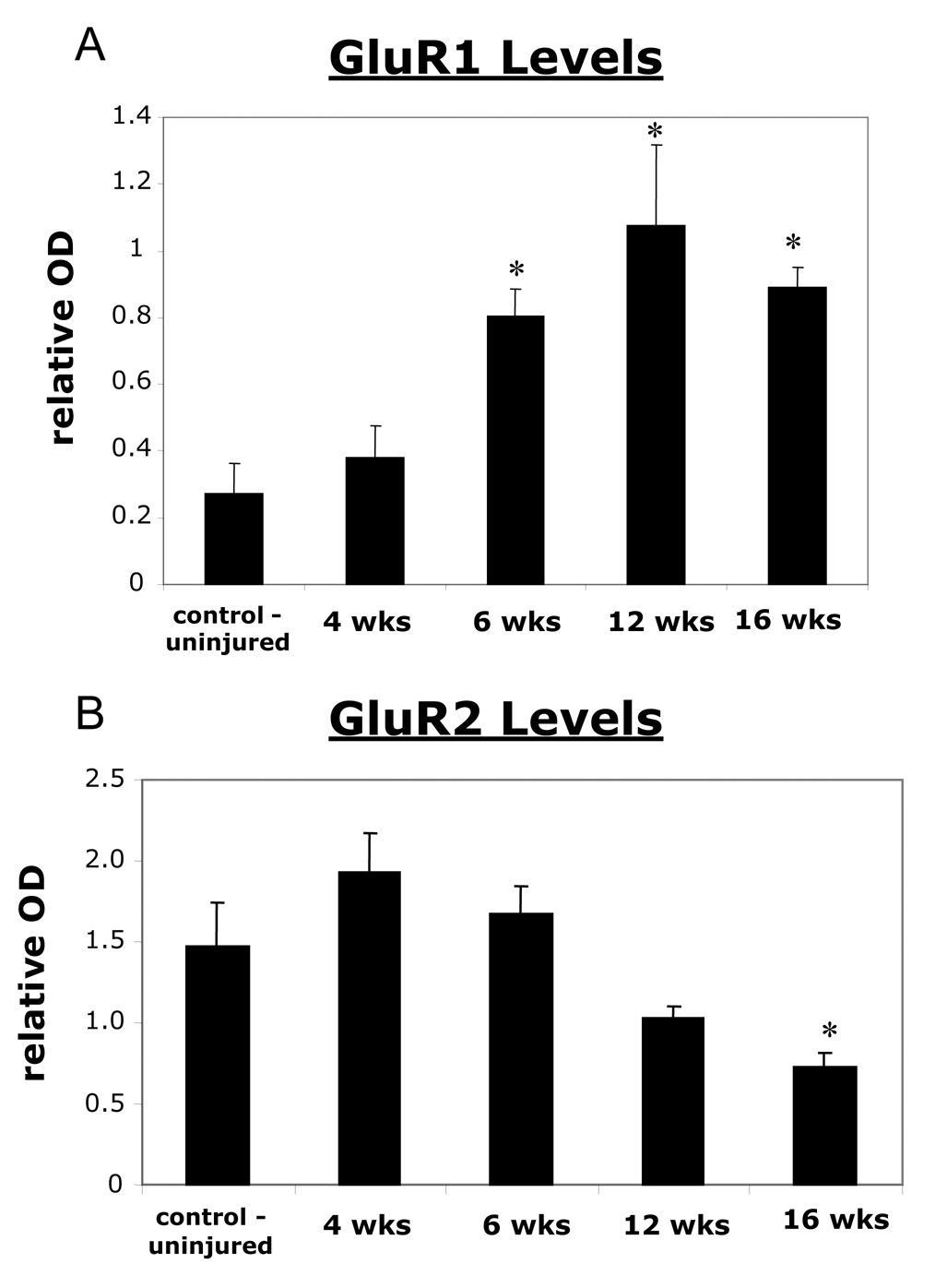
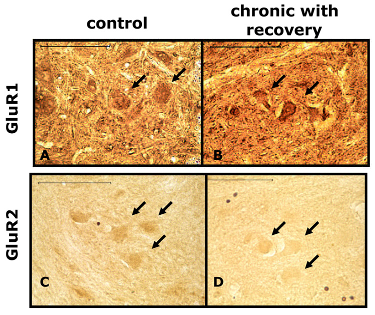
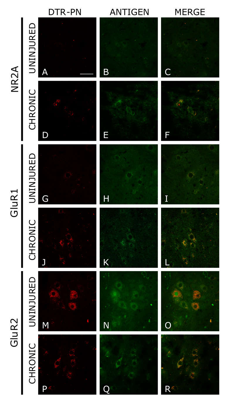
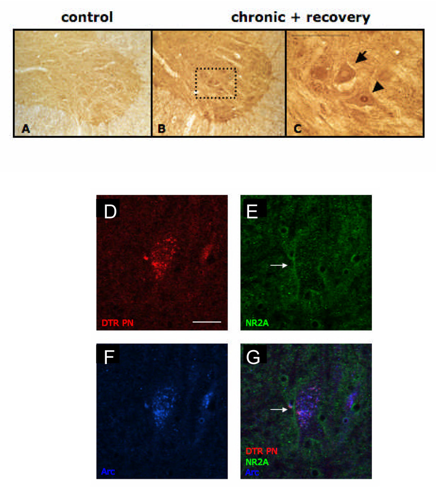
High cervical spinal cord hemisection results in paralysis of the ipsilateral hemidiaphragm; however, functional recovery of the paralyzed hemidiaphragm can occur spontaneously. The mechanisms mediating this recovery are unknown. In chronic, experimental contusive spinal cord injury, an upregulation of the NMDA receptor 2A subunit and a downregulation of the AMPA receptor GluR2 subunit have been correlated with improved hind limb motor recovery. Therefore, we hypothesized that NR2A is upregulated, whereas GluR2 is down-regulated following chronic C2 hemisection to initiate synaptic strengthening in respiratory motor pathways. Since NMDA receptor activation can lead to the delivery of AMPA receptor subunits to the post-synaptic membrane, we also hypothesized that there would be an upregulation of the GluR1 AMPA receptor subunit and that activity-regulated cytoskeletal associated protein may mediate the post-synaptic membrane delivery. Female rats were hemisected at C2 and allowed to recover for different time points following hemisection. At these time points, protein levels of NR2A, GluR1, and GluR2 subunits were assessed via Western blot analysis. Western blot analysis revealed that there were increases in NR2A subunit at six and twelve weeks post C2 hemisection. At six, twelve, and sixteen weeks post hemisection, the GluR1 subunit was increased over controls, whereas the GluR2 subunit decreased sixteen weeks post hemisection. Immunocytochemical data qualitatively supported these findings. Results also indicated that activity-regulated cytoskeletal associated protein may be associated with the above changes. These findings suggest a role of NR2A, GluR1, and GluR2 in mediating chronic spontaneous functional recovery of the paralyzed hemidiaphragm following cervical spinal cord hemisection.


Figures

Figure 1
Figure 1. Diagram of the crossed phrenic pathway
High cervical hemisection of the mammalian spinal cord rostral to the level of the phrenic nucleus interrupts (dotted lines) the descending bulbospinal respiratory drive to the ipsilateral phrenic nucleus, resulting in paralysis of the ipsilateral hemidiaphragm. Spontaneous activation of the latent pathway occurs as early as six weeks in 33% of hemisected animals of the present study. By 16 weeks, 67% of all C2 hemisected rats display recovery of the ipsilateral hemidiaphragm. The latent pathway descends contralateral to the hemisection and then crosses the spinal cord midline to innervate phrenic motor neurons ipsilateral to the lesion. The activation of these neurons induces recovery to the previously paralyzed hemidiaphragm.
Figure 2
Figure 2. Electromyograms from both the left and right sides of the diaphragm of an uninjured control rat and the same hemisected rat one week (acute) and 16 weeks (chronic) post injury
In the non-injured animal (traces on the left), the EMG recording from the left and right hemidiaphragm shows a rhythmic, synchronized activity. Acutely following left C2 hemisection (middle traces), the left hemidiaphragm displayed no EMG activity. Sixteen weeks following left C2 hemisection (traces on the right); however, the previously paralyzed left hemidiaphragm displayed spontaneously recovered EMG activity. Each recording is seven seconds long.
Figure 3
Figure 3. A graph showing an increase in NR2A subunit at 6 and 12 weeks post hemisection as revealed by Western blot analysis
All values are relative to a standard cortical tissue run along side the samples. Six and twelve weeks after left C2 hemisection, the left spinal cord (C3–C6) had a significantly higher relative optical density (OD) value corresponding to NR2A protein levels compared to control animals. At sixteen weeks, NR2A protein levels returned to values not significantly higher than controls. * indicates significance with a p < 0.05.
Figure 4
Figure 4. Light micrographs of ventral horn motor neurons and phrenic motor neurons (identified through retrograde labeling with WGA-HRP) showing increased NR2A immunoreactivity (IR) following chronic C2 hemisection in rats
Immunocytochemistry revealed that there was an increase in the density of NR2A IR in ventral horn motor neurons and phrenic motor neurons in C2 hemisected rats compared to control. A and B are from a control, non-hemisected rat. C and D are from a chronic (12 weeks) C2 hemisected animal with recovery. B and D show retrogradely labeled PMNs visualized with TMB (arrows). A and C are adjacent sections to B and D, respectively, and show NR2A immunoreactive motor neurons (arrowheads) as well as phrenic motor neurons (arrows). The circle is the relative location of the phrenic motor nucleus derived from the adjacent sections. Scale bars = 100 µm.
Figure 5
Figure 5. 5A) A graph showing that there is a significant increase of the AMPA GluR1 subunit receptor at 6, 12, and 16 weeks post C2 hemisection following Western blot analysis
All values are relative to a standard cortical tissue run along side the samples. 5B) A graph showing that there is a down regulation of the AMPA GluR2 subunit which is significantly lower than control at 16 weeks post C2 hemisection. The graph represents GluR2 values quantified through Western blot analysis of control uninjured animals compared to hemisected animals at various time points following injury. All values are relative to a standard cortical tissue run along side the samples. Significant values are reached at sixteen weeks post hemisection. * indicates significance with a p < 0.05.
Figure 6
Figure 6. Light micrographs showing that phrenic motor neurons had increased GluR1 and decreased GluR2 immunoreactivity following chronic C2 hemisection
A and C are sections from control, non-hemisected rats. B and D are sections from chronic C2 hemisected rats that displayed recovery. A and B are sections processed for GluR1 immunocytochemistry. C and D are sections that have been processed for GluR2 immunocytochemistry. Arrows are pointing to immunoreactive phrenic motor neurons. Note that GluR1 is increased in chronically (12 weeks) injured rats, while GluR2 is decreased in these animals. Scale bars = 100 µm.
Figure 7
Figure 7. Immunofluorescent images depicting membrane bound NR2A, GluR1, and GluR2 on phrenic motor neurons retrogradely labeled with dextran Texas red
A, D, G, J, M, P show phrenic motor neurons labeled with dextran Texas red. The first two rows depict the levels of NR2A on these labeled PMNs. In C and F there appears to be an upregulation of membrane bound NR2A in chronic lesioned animals compared to uninjured animals. The middle two rows show the levels of GluR1 on both chronic lesioned and unlesioned animals. In I and L, membrane bound GluR1 appears to be increased on PMNs. Magnification of all figures is the same and scale bar equals 50 um.
Figure 8
Figure 8. Arc was observed on ventral horn motor neurons following chronic C2 hemisection and was localized on both the cell body and nucleus
A and B are sections processed for Arc immunoreactivity. A is from a control, non-hemisected rat. B is from a chronic (12 weeks) C2 hemisected rat with spontaneous recovery. In a higher magnification photomicrograph of B (C), it is notable that Arc was localized to both the cell body (arrow) as well as the motor neuron nucleus (arrowhead). Scale bars = 100 µm. D–G) Triple label fluorescent micrographs showing that Arc and NR2A are both expressed in phrenic motor neurons. D is a picture of retrogradely labeled phrenic motor neurons (red). E and F are pictures of NR2A (green) and Arc (blue), respectively. G is an overlay of all three images. Magnification of all immunofluorescent figures is the same and scale bar equals 50 um.

References

    1. Alilain WJ, Goshgarian HG. MK-801 upregulates NR2A protein levels and induces functional recovery of the ipsilateral hemidiaphragm following acute C2 hemisection in adult rats. J Spinal Cord Med. 2007;30:346–354. - PMC - PubMed
    1. Alsbo CW, Wrang ML, Moller F, Diemer NH. Is the AMPA receptor subunit GluR2 mRNA an early indicator of cell fate after ischemia? A quantitative single cell RT-PCR study. Brain Res. 2001;894:101–108. - PubMed
    1. Asztely F, Erdemli G, Kullmann DM. Extrasynaptic glutamate spillover in the hippocampus: dependence on temperature and the role of active glutamate uptake. Neuron. 1997;18:281–293. - PubMed
    1. Barbour B, Hausser M. Intersynaptic diffusion of neurotransmitter. Trends Neurosci. 1997;20:377–384. - PubMed
    1. Bennett MV, Pellegrini-Giampietro DE, Gorter JA, Aronica E, Connor JA, Zukin RS. The GluR2 hypothesis: Ca(++)-permeable AMPA receptors in delayed neurodegeneration. Cold Spring Harb Symp Quant Biol. 1996;61:373–384. - PubMed

Publication types

MeSH terms

Substances