. 2008 Aug;160(2):107-15.
doi: 10.1016/j.molbiopara.2008.04.011. Epub 2008 May 2.

N-terminal processing of proteins exported by malaria parasites

Affiliations

N-terminal processing of proteins exported by malaria parasites

Henry H Chang et al. Mol Biochem Parasitol. 2008 Aug.

Abstract

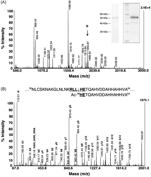
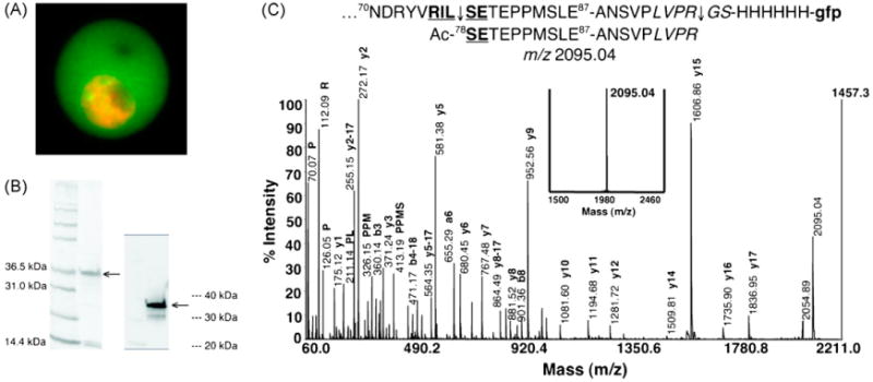
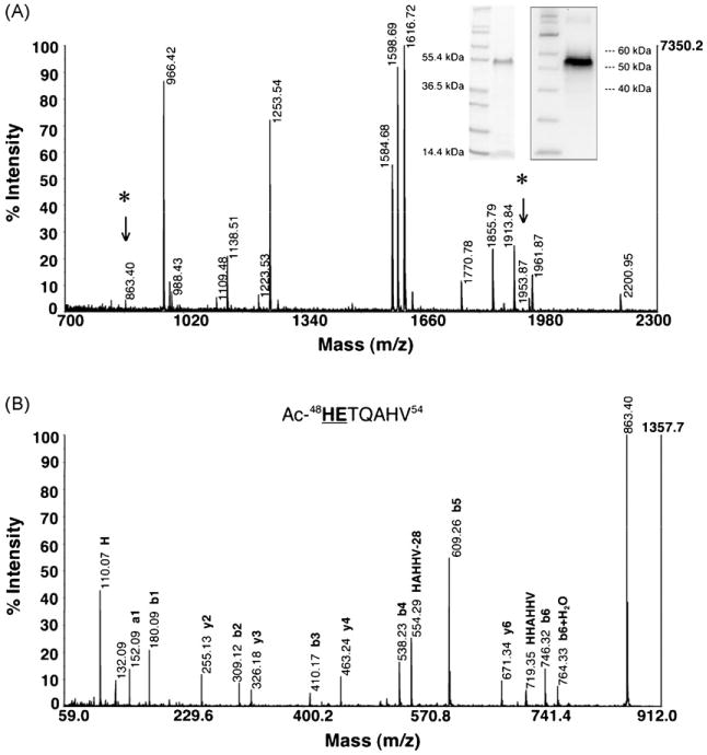
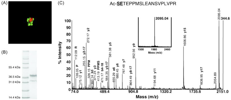
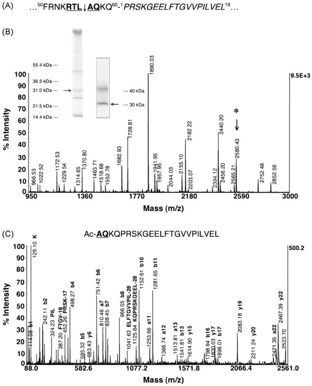
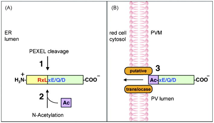
Malaria parasites utilize a short N-terminal amino acid motif termed the Plasmodium export element (PEXEL) to export an array of proteins to the host erythrocyte during blood stage infection. Using immunoaffinity chromatography and mass spectrometry, insight into this signal-mediated trafficking mechanism was gained by discovering that the PEXEL motif is cleaved and N-acetylated. PfHRPII and PfEMP2 are two soluble proteins exported by Plasmodium falciparum that were demonstrated to undergo PEXEL cleavage and N-acetylation, thus indicating that this N-terminal processing may be general to many exported soluble proteins. It was established that PEXEL processing occurs upstream of the brefeldin A-sensitive trafficking step in the P. falciparum secretory pathway, therefore cleavage and N-acetylation of the PEXEL motif occurs in the endoplasmic reticulum (ER) of the parasite. Furthermore, it was shown that the recognition of the processed N-terminus of exported proteins within the parasitophorous vacuole may be crucial for protein transport to the host erythrocyte. It appears that the PEXEL may be defined as a novel ER peptidase cleavage site and a classical N-acetyltransferase substrate sequence.


Figures

Fig. 1
Fig. 1
PEXEL of PfHRPII is cleaved and N-acetylated. (A) Native PfHRPII purified from the saponin supernatant of trophozoite-infected RBCs was subjected to an Asp-N solution digestion and analyzed by MALDI-TOF MS. The inset Coomassie-stained gel (left) and anti-PfHRPII Western blot (right) confirms the purified protein. The peptide fragment at m/z 1953.87 (*) was selected for tandem MS analysis. (B) De novo sequencing of PfHRPII m/z 1953.87. CID analysis reveals that m/z 1953.87 is the most N-terminal peptide fragment and demonstrates the PEXEL in PfHRPII (bold and underlined) is cleaved after residue 47 (45RLL↓) and the mature N-terminus is N-acetylated (Ac-HE49). The absolute intensity of the highest signal for each spectrum is labeled in bold on the right vertical axis (2.4E+4 and 1675.1, respectively).
Fig. 2
Fig. 2
PEXEL of PfEMP2 is cleaved and N-acetylated. (A) PfEMP2-GFP is exported to the host RBC. PfEMP2-GFP (green) was imaged live and the nuclei of the parasites were stained with DRAQ5 (orange). (B) PfEMP2-GFP was enriched from trophozoite-infected RBC saponin supernatant. The Coomassie-stained gel (left) and anti-GFP Western blot (right) confirms the enriched protein (arrows). The contaminating proteins at ∼15 kDa are likely the α and β subunits of hemoglobin. (C) PfEMP2-GFP was treated with thrombin to generate a single peptide at m/z 2095.04. The PfEMP2-GFP chimera has the first 87 amino acids of PfEMP2 containing the PEXEL 75RILSE79 (bold and underlined), a thrombin cleavage motif (italicized), and a His6 linker fused to the N-terminus of the GFP reporter (lower case). A peptide with a predicted m/z 2095.04 is expected to be produced after PEXEL processing and thrombin cleavage. Upon treatment of the enriched PfEMP2-GFP with thrombin, a single peptide at m/z 2095.04 was indeed generated (bold inset spectrum). Tandem MS analysis by CID produced a fragmentation pattern that confirms the sequence of the generated peptide to reflect the cleavage (75RIL↓) and N-acetylation (Ac-SE79) of the PEXEL in PfEMP2.
Fig. 3
Fig. 3
PfHRPIImyc PEXEL is processed upstream of the BFA-sensitive trafficking step. (A) PfHRPIImyc trapped in the ER from BFA treatment was extracted and enriched from the resulting parasite pellet after saponin lysis. The inset Coomassie-stained gel (left) and anti-c-Myc Western blot (right) confirms the enriched protein. An Asp-N in-gel digestion was performed because of contaminating hemoglobin (∼15 kDa). After accounting for all predicted Asp-N proteolysis products, the peptide fragments at m/z 863.40 (*) and 1953.87 (*) were selected for tandem MS analysis. (B) De novo sequencing of PfHRPIImyc m/z 863.40. CID analysis reveals that m/z 863.40 is one of the N-terminal peptide fragments along with m/z 1953.87 (not shown) to demonstrate that the PEXEL processing of PfHRPIImyc occurs upstream of the BFA-sensitive trafficking step. Residues in the sequenced peptide that are bold and underlined are a part of the PfHRPIImyc PEXEL.
Fig. 4
Fig. 4
PEXEL processing occurs in the parasite ER. (A) Transfected parasites expressing PfEMP2-GFP were treated with 5 μg/mL BFA for 24 h. Live imaging of the BFA-treated parasites indicates that the trafficking of PfEMP2-GFP (green) is BFA-sensitive. Since the parasite nucleus is stained with DRAQ5 (orange), chimeric protein is exclusively localized to a perinuclear compartment indicative of the parasite ER. (B) PfEMP2-GFP trapped in the ER from BFA treatment was enriched from the parasite pellet after saponin lysis. The predominant upper band at ∼35 kDa is the full-length chimera while the lower band at ∼30 kDa is a GFP degradation product seen previously in the anti-GFP Western blot in Fig. 2B. (C) Thrombin cleavage of the ER-trapped PfEMP2-GFP produced a single peptide at m/z 2095.04 (bold inset spectrum). This peptide was confirmed by CID to correspond to the fragment generated after PEXEL processing and thrombin cleavage. The cleavage and N-acetylation of the PEXEL prior to anterograde transport between the ER and Golgi reveals that PEXEL processing occurs in the parasite ER.
Fig. 5
Fig. 5
Transport across the PVM may be mediated by the recognition of the processed PEXEL. (A) KAHRP(1-60)-GFP traffics to the PV lumen despite having a full PEXEL motif (bold and underlined) because of its close proximity to the N-terminus of GFP (italicized). (B) KAHRP(1-60)-GFP trapped in the PV of trophozoite parasites was enriched from saponin supernatant and subjected to an Asp-N in-gel digestion. The inset Coomassie-stained gel (left) and anti-GFP Western blot (right) confirms the enriched protein (arrows) at ∼31 kDa while the upper band (∼40 kDa) is a cross-reactive protein. The peptide fragment at m/z 2580.43 (*) was selected for CID analysis. (C) De novo sequencing of m/z 2580.43 by CID establishes that the PEXEL of KAHRP(1-60)-GFP is cleaved (54RTL↓) and N-acetylated (Ac-AQ58), thus indicating that PEXEL processing in the ER is not affected by the close proximity of the GFP reporter to the PEXEL, but there may be a failure in the recognition of the processed PEXEL (Ac-xE/Q/D) by a putative translocase at the PVM.
Fig. 6
Fig. 6
Model illustrating PEXEL processing and the definition of the PEXEL. (Panel A) Soluble proteins entering the P. falciparum secretory pathway undergo PEXEL cleavage (1) and N-acetylation (2) in the parasite ER lumen. (Panel B) The cleaved, N-acetylated protein is recognized by the putative translocase at the PVM (3) for export to the host RBC. The PEXEL may be defined as a novel ER peptidase cleavage site (RxL) (red) and a classical N-acetyltransferase substrate sequence (xE/Q/D) (blue).

References

    1. Miller LH, Baruch DI, Marsh K, Doumbo OK. The pathogenic basis of malaria. Nature. 2002;415:673–9. - PubMed
    1. Kirk K. Membrane transport in the malaria-infected erythrocyte. Physiol Rev. 2001;81:495–537. - PubMed
    1. Kilejian A. Characterization of a protein correlated with the production of knob-like protrusions on membranes of erythrocytes infected with Plasmodium falciparum. Proc Natl Acad Sci USA. 1979;76:4650–3. - PMC - PubMed
    1. Pologe LG, Ravetch JV. A chromosomal rearrangement in a P. falciparum histidine-rich protein gene is associated with the knobless phenotype. Nature. 1986;322:474–7. - PubMed
    1. Baruch DI, Pasloske BL, Singh HB, et al. Cloning the P. falciparum gene encoding PfEMP1, a malarial variant antigen and adherence receptor on the surface of parasitized human erythrocytes. Cell. 1995;82:77–87. - PubMed

Publication types

LinkOut - more resources