. 2008 Aug 8;283(32):22076-88.
doi: 10.1074/jbc.M803901200. Epub 2008 Jun 4.

Hhat is a palmitoylacyltransferase with specificity for N-palmitoylation of Sonic Hedgehog

Affiliations

Hhat is a palmitoylacyltransferase with specificity for N-palmitoylation of Sonic Hedgehog

John A Buglino et al. J Biol Chem. .

Abstract

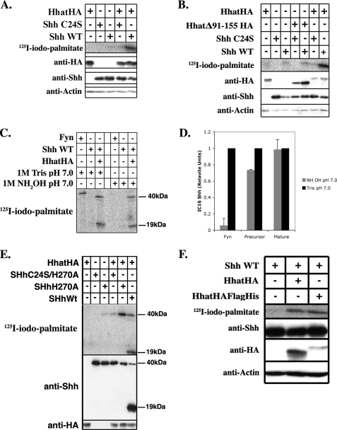
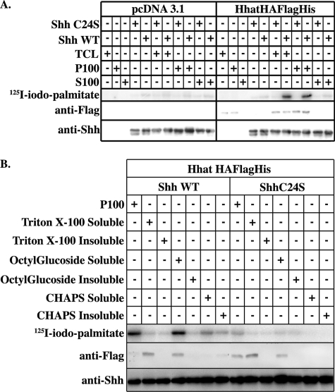
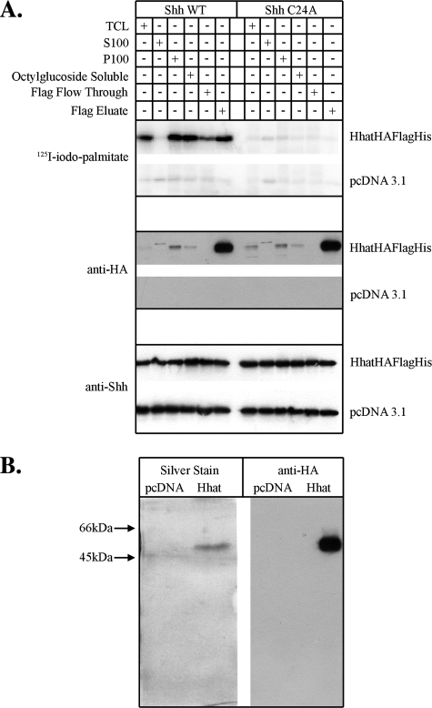
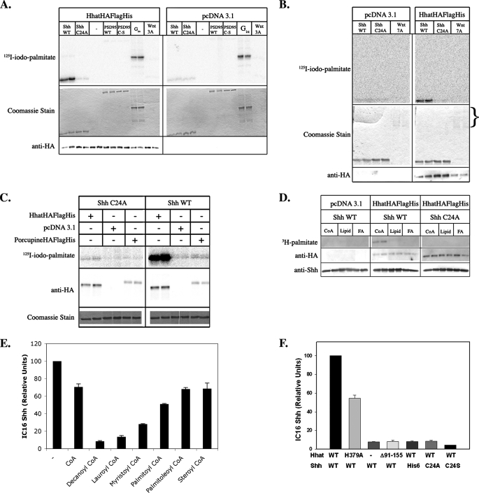
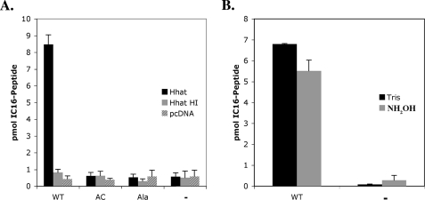
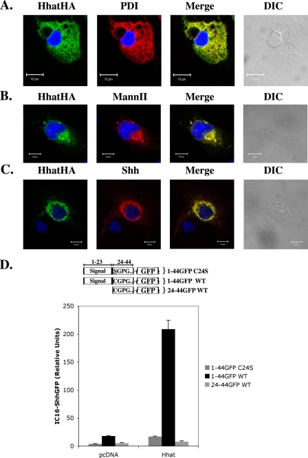
Palmitoylation of Sonic Hedgehog (Shh) is critical for effective long- and short-range signaling. Genetic screens uncovered a potential palmitoylacyltransferase (PAT) for Shh, Hhat, but the molecular mechanism of Shh palmitoylation remains unclear. Here, we have developed and exploited an in vitro Shh palmitoylation assay to purify Hhat to homogeneity. We provide direct biochemical evidence that Hhat is a PAT with specificity for attaching palmitate via amide linkage to the N-terminal cysteine of Shh. Other palmitoylated proteins (e.g. PSD95 and Wnt) are not substrates for Hhat, and Porcupine, a putative Wnt PAT, does not palmitoylate Shh. Neither autocleavage nor cholesterol modification is required for Shh palmitoylation. Both the Shh precursor and mature protein are N-palmitoylated by Hhat, and the reaction occurs during passage through the secretory pathway. This study establishes Hhat as a bona fide Shh PAT and serves as a model for understanding how secreted morphogens are modified by distinct PATs.


Figures

FIGURE 1.
FIGURE 1.
Reconstitution of Shh palmitoylation in vivo. COS-1 cells were transfected with the indicated constructs and labeled with [125I]iodopalmitate for 4 h. Cell lysates were analyzed directly by Western blotting or after Shh immunoprecipitation. A, B, E, and F: upper panels, [125I]iodopalmitate incorporation into immunoprecipitated Shh as detected by phosphorimaging; lower panels, Western blots of the same extracts probed with anti-HA, anti-Shh, and anti-actin antibodies. C: immunoprecipitated samples containing [125I]iodopalmitate-labeled Fyn or Shh loaded in duplicate on a 12.5% SDS-polyacrylamide gel. After electrophoresis, the gel was split in half. Each half was incubated in either 1 m Tris or NH2OH for 1 h at 20°C and then analyzed by phosphorimaging. D: quantification of the experiment in C, performed three times. Phosphorimaging signals were normalized for levels of protein expression; NH2OH sensitivity is expressed as a percentage of Tris-treated controls. WT, wild-type.
FIGURE 2.
FIGURE 2.
Partially purified Hhat-HA-FLAG-His stimulates Shh palmitoylation in vitro. A, COS-1 cells transfected with Hhat-HA-FLAG-His or empty pcDNA3.1 vector were lysed and separated into S100 and P100 fraction as described under “Experimental Procedures.” 10 μl of each fraction was reacted with 2 μg of recombinant wild-type (WT) Shh or Shh C24A and 100 μm [125I]iodopalmitoyl-CoA in reaction buffer (20 mm HEPES (pH 7.3), 200 mm NaCl, 1 mm ATP, 1 mm MgCl2, 1 mm DTT, and 0.05% Triton X-100) for 1 h at room temperature and then separated on 12.5% SDS-polyacrylamide gels. TCL, total cell lysate. B, P100 membranes derived from Hhat-HA-FLAG-His- or pcDNA3.1-expressing cells, as described above, were solubilized in buffer containing 1% octyl glucoside, Triton X-100, or CHAPS. Following ultracentrifugation, detergent-soluble and -insoluble fractions were tested for PAT activity as described above. In A and B, the upper panels are phosphorimages showing [125I]iodopalmitate incorporation into Shh. The lower panels are Western blots of the same samples probed with anti-FLAG and anti-Shh antibodies.
FIGURE 3.
FIGURE 3.
Purification of Hhat to homogeneity. A, 293FT cells transfected with Hhat-HA-FLAG-His or pcDNA3.1 vector were lysed, and Hhat-HA-FLAG-His was purified as described under “Experimental Procedures.” 10 μl of the indicated fraction was assayed for PAT activity as described in the legend to Fig. 2. Upper panels, phosphorimages showing [125I]iodopalmitate incorporation into Shh; middle and lower panels, Western blots of the same samples probed with anti-HA and anti-Shh antibodies. WT, wild-type; TCL, total cell lysate. B, shown are silver stain and Western blot analysis of purified Hhat-HA-FLAG-His. The FLAG eluate fraction was concentrated 5-fold and then electrophoresed on a 12.5% SDS-polyacrylamide gel. Gels were either fixed and silver-stained or Western-blotted with Anti-HA antibodies. The entire length of each gel is shown.
FIGURE 4.
FIGURE 4.
Characterization of Hhat PAT activity. A, purified Shh (70 pmol) was incubated with purified Hhat-HA-FLAG-His (2. 5 pmol) or FLAG eluate from mock-transfected samples in the presence of 100 μm [125I]iodopalmitoyl-CoA for the indicated time points.B, the 1-h time point reaction was analyzed by SDS-PAGE, and gels were incubated in either 1 m Tris or NH2OH for 1 h and dried. Phosphorimaging signals were normalized for protein expression levels; NH2OH sensitivity is expressed as a percentage of Tris-treated controls. C and D, the reaction was performed for 1 h at the indicated NaCl concentrations or pH, respectively. E, Shh at the indicated concentrations was incubated with purified Hhat-HA-FLAG-His (2.5 pmol) in the presence of 100 μm [125I]iodopalmitoyl-CoA for 1 h. F, [125I]iodopalmitoyl-CoA at the indicated concentrations was incubated with purified Hhat-HA-FLAG-His (2.5 pmol) in the presence of 50 μm Shh. The insets for E and F represent Lineweaver-Burk plots. For each panel, Shh protein bands were excised from dried gels, and the amount of [125I]iodopalmitate incorporation was determined by γ-counting. Graphs represent the average of three experiments corrected for nonspecific incorporation of [125I]iodopalmitate as described under “Experimental Procedures.”
FIGURE 5.
FIGURE 5.
Hhat PAT activity is specific for Shh. A and B, purified Hhat-HA-FLAG-His (2. 5 pmol) was incubated either alone (-) or with 2 μg of wild-type (WT) Shh, Shh C24A, wild-type PSD95, PSD95 Cys → Ser, Gαi, or Wnt7A or 0.2 μg or Wnt3A and 100 μm [125I]iodopalmitoyl-CoA for 1 h at room temperature. [125I]Iodopalmitate incorporation was detected by phosphorimaging (upper panels). Substrate proteins were detected by Coomassie Blue staining (middle panels). Hhat-HA-FLAG-His levels were detected by Western blotting with anti-HA antibody (lower panels). Wnt proteins are glycosylated at multiple sites, which makes analysis of Wnt7A levels by SDS-PAGE and Coomassie Blue staining problematic (note multiple diffuse bands). However, protein assays confirmed that Wnt7A was present at levels similar to Shh. C, P100 membranes derived from 293FT cells expressing Hhat-HA-FLAG-His, Porc-HA-FLAG-His, or empty pcDNA3.1 vector were incubated with 2 μg of wild-type Shh or Shh C24A along with 100 μm [125I]iodopalmitoyl-CoA for 1 h at room temperature. Detection was performed as described above. D, purified Hhat-HA-FLAG-His (2.5 pmol) was incubated with 2 μg of wild-type Shh or Shh C24A along with 20 μm either [3H]palmitoyl-CoA (CoA), [3H]dipalmitoylphosphatidylethanolamine (Lipid), or [3H]palmitic acid (FA) for 1 h at room temperature. [3H]Palmitate incorporation was detected by fluorography (upper panel). Hhat-HA-FLAG-His and Shh were detected by Western blotting with anti-HA antibody (middle panel) or anti-Shh antibody (lower panel). E, purified Hhat-HA-FLAG-His (2.5 pmol) was incubated with 2 μg of purified wild-type Shh and 50 μm [125I]iodopalmitoyl-CoA alone (-) or with the indicated CoA (100 μm) for 1 h at room temperature. [125I]Iodopalmitate incorporation was detected by phosphorimaging (IC16-Shh). Graphs represent the average of three experiments. F, 10 μl (2.5 pmol) of purified Hhat-FLAG-His (WT), Hhat-FLAG-His-H379A (H379A), Hhat-HA-His-FLAGΔ91–155 (Δ91–155), or FLAG eluate from mock-transfected samples (-) was incubated with 2 μg of wild-type Shh, Shh C24A, Shh C24S, or N-terminally His6-tagged wild-type Shh (His6) for 1 h at room temperature. [125I]Iodopalmitate incorporation was detected by phosphorimaging (IC16-Shh). Graphs represent the average of three experiments.
FIGURE 6.
FIGURE 6.
A peptide containing the first 11 amino acids of Shh is palmitoylated by Hhat. A, 10 μl (5 pmol) of purified Hhat-HA-FLAG-His that was untreated (Hhat) or heat-inactivated (95 °C, 5 min; Hhat HI) or FLAG eluate from mock-transfected samples (pcDNA) was incubated either alone (-) or with 100 μm wild-type biotinylated Shh peptide (WT), N-terminally acetylated biotinylated Shh peptide (AC), or biotinylated Shh peptide (Ala) for 1 h at room temperature. Biotinylated peptides were precipitated using streptavidin-agarose. [125I]Iodopalmitate incorporation was determined byγ-counting. B, hydroxylamine sensitivity was determined by soaking wild-type and no-substrate (-) samples in 0.1 m Tris or NH2OH for 18 h at room temperature. Graphs represent the average of three experiments.
FIGURE 7.
FIGURE 7.
Hhat-mediated Shh palmitoylation occurs within the secretory pathway. A–C, COS-1 cells transfected with Hhat-HA alone or with full-length Shh were fixed and processed for indirect immunofluorescence. Hhat-HA colocalization with protein-disulfide isomerase (PDI) (A), mannosidase II (MannII) (B), and Shh (C) is depicted. D, COS-1 cells cotransfected with the indicated Shh-GFP fusion protein (schematized above graph) and either Hhat-HA or empty pcDNA3.1 vector were labeled with [125I]iodopalmitate. Fusion proteins were immunoprecipitated with anti-GFP antibody, and [125I]iodopalmitate incorporation was detected by phosphorimaging. Graphs represent the average of three experiments. WT, wild-type.

References

    1. McMahon, A. P., Ingham, P. W., and Tabin, C. J. (2003) Curr. Top. Dev. Biol. 53 1-114 - PubMed
    1. Fuccillo, M., Joyner, A. L., and Fishell, G. (2006) Nat. Rev. Neurosci. 7 772-783 - PubMed
    1. Ho, K. S., and Scott, M. P. (2002) Curr. Opin. Neurobiol. 12 57-63 - PubMed
    1. Chiang, C., Litingtung, Y., Lee, E., Young, K. E., Corden, J. L., Westphal, H., and Beachy, P. A. (1996) Nature 383 407-413 - PubMed
    1. Roessler, E., Belloni, E., Gaudenz, K., Vargas, F., Scherer, S. W., Tsui, L. C., and Muenke, M. (1997) Hum. Mol. Genet 6 1847-1853 - PubMed

Publication types