Review
. 2008 Jun;72(2):249-65, table of contents.
doi: 10.1128/MMBR.00040-07.

Tegument proteins of human cytomegalovirus

Affiliations
Review

Tegument proteins of human cytomegalovirus

Robert F Kalejta. Microbiol Mol Biol Rev. 2008 Jun.

Abstract

Human cytomegalovirus (HCMV) is a common, medically relevant human herpesvirus. The tegument layer of herpesvirus virions lies between the genome-containing capsids and the viral envelope. Proteins within the tegument layer of herpesviruses are released into the cell upon entry when the viral envelope fuses with the cell membrane. These proteins are fully formed and active and control viral entry, gene expression, and immune evasion. Most tegument proteins accumulate to high levels during later stages of infection, when they direct the assembly and egress of progeny virions. Thus, viral tegument proteins play critical roles at the very earliest and very last steps of the HCMV lytic replication cycle. This review summarizes HCMV tegument composition and structure as well as the known and speculated functions of viral tegument proteins. Important directions for future investigation and the challenges that lie ahead are identified and discussed.


Figures

FIG. 1.
FIG. 1.
The HCMV virion. The cartoon represents (not to scale) an average HCMV infectious viral particle. Abundant tegument proteins are listed. The large shapes on the surface of the virion represent various virally encoded membrane glycoproteins. Please see the text for further details.
FIG. 2.
FIG. 2.
Tegument proteins help deliver the HCMV genome-containing capsid to the nucleus during the viral entry process. (A) Signals initiated upon receptor binding induce cellular antiviral responses but may also prime the cell for subsequent events during viral entry. (B) Capsid-associated tegument proteins UL47 and UL48 (and perhaps pp150) direct capsids along microtubules (MTs) toward nuclear pore complexes. Cellular motor proteins such as dynein (not shown) likely assist this transport. (C) A subset of tegument proteins (such as pp65 and pp71) is transported to the nucleus independently of capsids. (D) Capsids eventually dissociate from microtubules, dock at nuclear pores, and release their DNA into the nucleus. The role of tegument proteins in this process is implicated but has not been described in detail.
FIG. 3.
FIG. 3.
Subcellular localization of tegument-delivered pp71 determines whether HCMV initiates lytic replication or establishes quiescent, latent-like infections. (A) Lytic replication initiates when tegument-delivered pp71 is allowed access to the nucleus. Capsids docked at nuclear pores release their DNA into the nucleus, and viral genomes associate with cellular histones (H). The Daxx protein, which rapidly dissociates from, and reassociates with, PML-NBs, accumulates around viral genomes, recruits an HDAC, and silences viral IE gene expression. Other PML-NB components are also recruited and participate in the silencing of viral genomes. pp71 binds to Daxx in these newly formed PML-NBs, induces Daxx degradation, derepresses viral IE gene expression, and thus initiates the lytic replication cycle. (B) In cells where quiescent or latent infections are established, tegument-delivered pp71 remains in the cytoplasm. Daxx (and presumably other PML-NB proteins) silences viral gene expression in these cells.
FIG. 4.
FIG. 4.
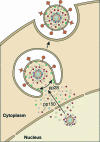
HCMV egress. DNA-containing capsids may associate with some tegument proteins while still in the nucleus. After exiting the nucleus by an envelopment-deenvelopment pathway (not shown), they acquire more tegument proteins in the cytoplasm. Tegumented capsids migrate to assembly sites located on Golgi apparatus-derived vesicles, where they obtain their final envelope that contains viral glycoproteins. The eventual fusion of these vesicles with the cell membrane results in the release of fully formed virions. The pp150 protein likely plays a role in directing capsids to assembly sites, and the pp28 protein likely plays a role in the formation of viral particles. Symbols are as shown in Fig. 1.

Similar articles

Cited by

References

    1. Abate, D. A., S. Watanabe, and E. Mocarski. 2004. Major human cytomegalovirus structural protein pp65 (ppUL83) prevents interferon response factor 3 activation in the interferon response. J. Virol. 7810995-11006. - PMC - PubMed
    1. Adair, R., E. R. Douglas, J. B. Maclean, S. Y. Graham, J. D. Aitken, F. E. Jamieson, and D. J. Dargan. 2002. The products of human cytomegalovirus genes UL23, UL24, UL43 and US22 are tegument components. J. Gen. Virol. 831315-1324. - PubMed
    1. Adamo, J. E., J. Schroer, and T. Shenk. 2004. Human cytomegalovirus TRS1 protein is required for efficient assembly of DNA-containing capsids. J. Virol. 7810221-10229. - PMC - PubMed
    1. Agromayor, M., and J. Martin-Serrano. 2006. Interaction of AMSH with ESCRT-III and deubiquitination of endosomal cargo. J. Biol. Chem. 28123083-23091. - PubMed
    1. Andoniou, C. E., and M. A. Degli-Esposti. 2006. Insights into the mechanisms of CMV-mediated interference with cellular apoptosis. Immunol. Cell Biol. 8499-106. - PubMed

Publication types

Substances

LinkOut - more resources