. 2008 Jun;72(2):301-16, table of contents.
doi: 10.1128/MMBR.00038-07.

Structural biology of pectin degradation by Enterobacteriaceae

Affiliations

Structural biology of pectin degradation by Enterobacteriaceae

D Wade Abbott et al. Microbiol Mol Biol Rev. 2008 Jun.

Abstract

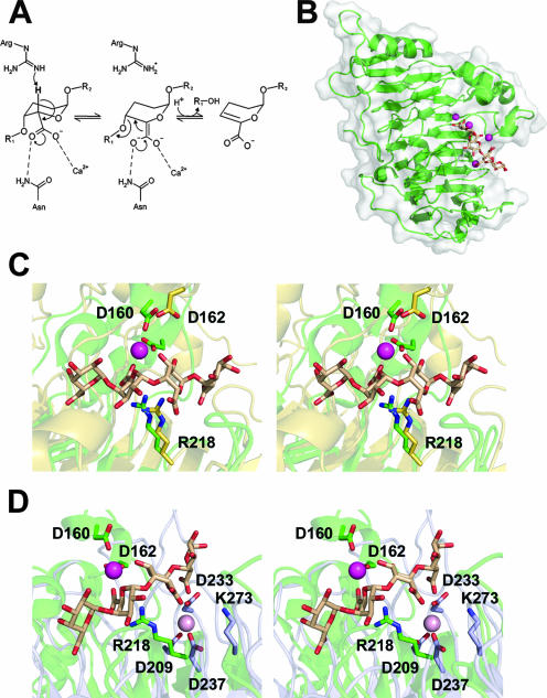
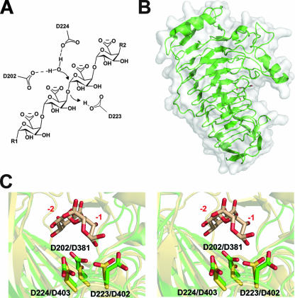
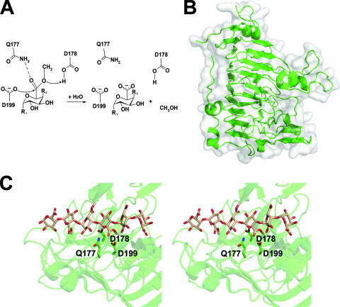
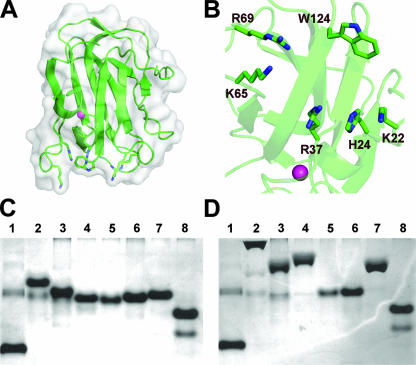
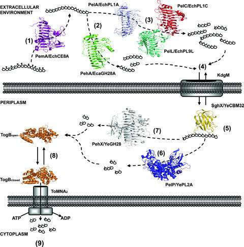
Pectin is a structural polysaccharide that is integral for the stability of plant cell walls. During soft rot infection, secreted virulence factors from pectinolytic bacteria such as Erwinia spp. degrade pectin, resulting in characteristic plant cell necrosis and tissue maceration. Catabolism of pectin and its breakdown products by pectinolytic bacteria occurs within distinct cellular environments. This process initiates outside the cell, continues within the periplasmic space, and culminates in the cytoplasm. Although pectin utilization is well understood at the genetic and biochemical levels, an inclusive structural description of pectinases and pectin binding proteins by both extracellular and periplasmic enzymes has been lacking, especially following the recent characterization of several periplasmic components and protein-oligogalacturonide complexes. Here we provide a comprehensive analysis of the protein folds and mechanisms of pectate lyases, polygalacturonases, and carbohydrate esterases and the binding specificities of two periplasmic pectic binding proteins from Enterobacteriaceae. This review provides a structural understanding of the molecular determinants of pectin utilization and the mechanisms driving catabolite selectivity and flow through the pathway.


Figures

FIG. 1.
FIG. 1.
The extracellular endo-Pels PelA/EchPL1A, PelC/EchPL1C, and PelL/EchPL9A. (A) A generalized reaction coordinate for calcium assisted β-elimination. (B) PelC/EchPL1C (PDB ID, 2EWE) is displayed in a “cartoon” format with a transparent solvent-accessible surface. The pentagalacturonate substrate is shown as sticks colored beige and the calcium ions as spheres colored mageneta. (C) The superimposed active sites of PelC/EchPL1C (green) and PelA/EchPL1A (yellow) (PDB ID, 1OOC) displayed in a wall-eyed format. The structurally conserved Brønstead base and calcium-coordinating aspartate residues are shown as sticks and labeled with PelC/EchPL1C numbering. (D) The superimposed active sites of PelC/EchPL1C (green) and PelL/EchPL9A (blue) based upon overall enzyme alignment displayed in stereo. The catalytic base, a lysine within PL family 9, and calcium ion (pink) are structurally conserved but shifted laterally toward the reducing end of the superimposed substrate.
FIG. 2.
FIG. 2.
The extracellular endopolygalacturonase PehA/EcaGH28A. (A) Generalized reaction mechanism for inverting family 28 GHs. (Based on data from reference .) (B) PehA/EcaGH28A (PDB ID, 1BHE) is displayed in a “cartoon” format with a transparent solvent-accessible surface. Catalytic residues are displayed as sticks. (C) Superimposed catalytic sites of the closely related endopolygalacturonase PelA/EcaGH28A (green) and periplasmic exopolygalacturonase PehX/YeGH28 (yellow) (PDB ID, 2UVF) displayed in wall-eyed format. The residues from both PehA/EcaGH28A (D202, D223, and D224) and PehX/YeGH28 (D381, D402, and D403) are labeled. The digalacturonate product from the exopolygalacturonase complex is shown in beige, and subsites −1 and −2 are labeled in red.
FIG. 3.
FIG. 3.
The extracellular Pem PemA/EchCE8. (A) Generalized mechanism for demethylation of pectin by Pems. The nucleophile (D199) attacks the carbonyl carbon, forming a tetrahedral intermediate that is stabilized by Q177. The general acid-base catalyst D178 protonates the ester-linked oxygen, and attack by a catalytic water releases methanol and polygalacturonate, recharging the active site. (B) PemA/EchCE8A (PDB ID, 1QJV) is displayed in a “cartoon” format with a transparent solvent-accessible surface. (C) The active site of PemA/EchCE8A displayed in wall-eyed stereo. The bound hexasaccharide substrate (compound II) from E. chrysanthemi (PDB ID, 2NST) is shown, and the catalytic residue D178 has been reintroduced for reference.
FIG. 4.
FIG. 4.
The periplasmic polygalacturonic acid binding protein SghX/YeCBM32. (A) SghX/YeCBM32 (PDB ID, 2JDA) is displayed in a “cartoon” format with a transparent solvent-accessible surface. The structural calcium is shown as a sphere in magenta. (B) Binding site of SghX/YeCBM32 displaying the basic amino acids potentially involved in ligand recognition. (C) Native acrylamide gel electrophoresis of SghX/YeCBM32 mutants. Mutant protein was produced as described previously (5, 6), and ∼5 μg of purified SghX/YeCBM32 protein was electrophoresed through 10% acrylamide gels in the presence and absence of 0.1% polygalacturonate purified from citrus fruit at 100 V for 3.5 h. Lanes: 1, bovine serum albumin control; 2, wild type; 3, ΔK22A; 4, ΔH24A; 5, ΔK22A/H24A; 6, ΔR37A; 7, ΔK65A; 8, ΔK65A/R69A. (D) Polygalacturonate acrylamide (0.1%) gel electrophoresis of SghX/YeCBM32 mutants. Lanes are loaded in the same order as in panel C.
FIG. 5.
FIG. 5.
The periplasmic endo-Pel PelP/YePL2A. (A) PelP/YePL2A (PDB ID, 2V8J) is displayed in a “cartoon” format with a transparent solvent-accessible surface. The catalytic Mn2+ is shown as a light blue sphere. (B) Superimposition of the metal (green) and trigalacturonate (yellow) (PDB ID, 2V8K) complexes. Enzymes are rendered in a ribbon format and the substrate as sticks in beige. (C) Superimposition of the active sites from family 2 PelP/YePL2A and family 1 PelC/EchPL1C Pels displayed in wall-eyed format. The Mn2+ coordination pocket from the metal complex has been introduced for reference. The Mn2+ is shown in blue and the Ca2+ from PelC/EchPL1C in magenta.
FIG. 6.
FIG. 6.
The periplasmic exopolygalacturonase PehX/YeGH28. (A) PehX/YeGH28 in complex with digalacturonate (PDB ID, 2UVF) is displayed in a “cartoon” format with a transparent solvent-accessible surface and the disaccharide in beige. (B) The active-site surface of the exopolygalacturonase is shown with its two accessible subsites (−1 and −2). The putative catalytic acid D402 is shown in red.
FIG. 7.
FIG. 7.
TogB, the periplasmic solute binding component of the oligogalacturonide transporter TogMNAB from Y. enterocolitica. (A) TogB (PDB ID, 2UVG) is displayed in a “cartoon” format with a transparent solvent-accessible surface. (B) TogB in complex with 4,5-unsaturated digalacturonate (PDB ID, 2UVI) displayed in a “cartoon” format with a transparent solvent-accessible surface in the same orientation as in panel A to demonstrate the large conformational change induced upon binding. (C) Superimposition on the ligands within the saturated (PDB ID, 2UVH) and 4,5-unsaturated digalacturonate complexes. The distances between the uronate groups and S271 are shown for the saturated ligand (3.8 Å) and the unsaturated ligand (2.7 Å).
FIG. 8.
FIG. 8.
Structural biology of pectin degradation and transport within Enterobacteriaceae. The major stages of extracellular and periplasmic pectin utilization are shown. (1) Methylated polygalacturonate is de-esterified by PemA/EchCE8A (violet). Methoxyl groups are indicated by open circles. Extracellular depolymerization reactions occur predominantly by endo-acting enzymes. These reactions can occur by either a hydrolysis mechanism as shown for PehA/EcaGH28A (green) (2) or β-elimination by the Pels PelC/EchPL1C (red), PelA/EchPL1A (light purple), and PelL/EchPL9A (teal) (3). (4) The products of these reactions enter the periplasm by facilitated diffusion through the porin KdgM. (5) Retention of substrates within the periplasm is facilitated by SghX/YeCBM32 (yellow). Periplasmic depolymerizations are catalyzed by the endo-Pel PelP/YePL2A (blue) (6) or exopolygalacturonase PehX/YeGH28 (gray) (7). Oligogalacturonate products are bound by the TogB periplasmic binding protein (orange) and directed to the TogMNA components of the ABC transporter (8), where they are shuttled across the inner membrane in an ATP-coupled reaction (9).

Similar articles

Cited by

References

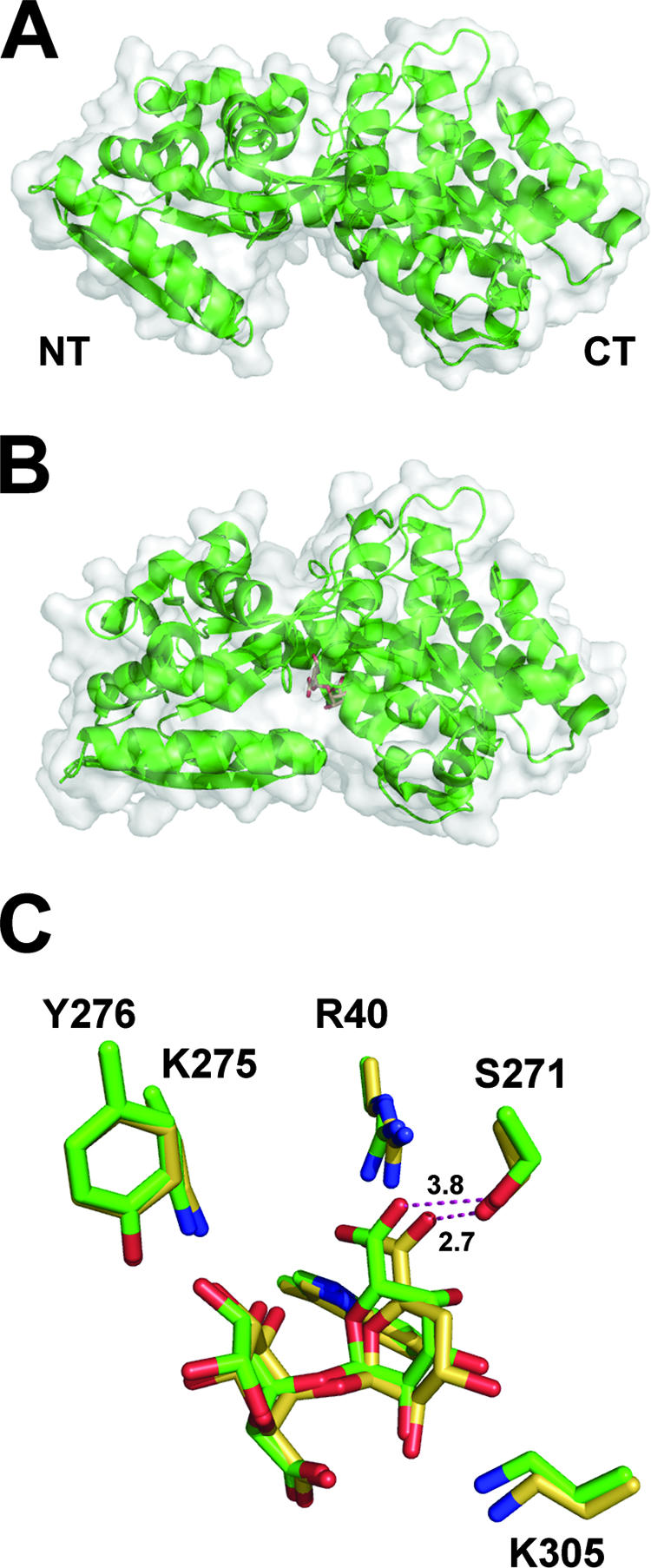
    1. Abbott, D. W., and A. B. Boraston. 2007. Specific recognition of saturated and 4,5-unsaturated hexuronate sugars by a periplasmic binding protein involved in pectin catabolism. J. Mol. Biol. 369759-770. - PubMed
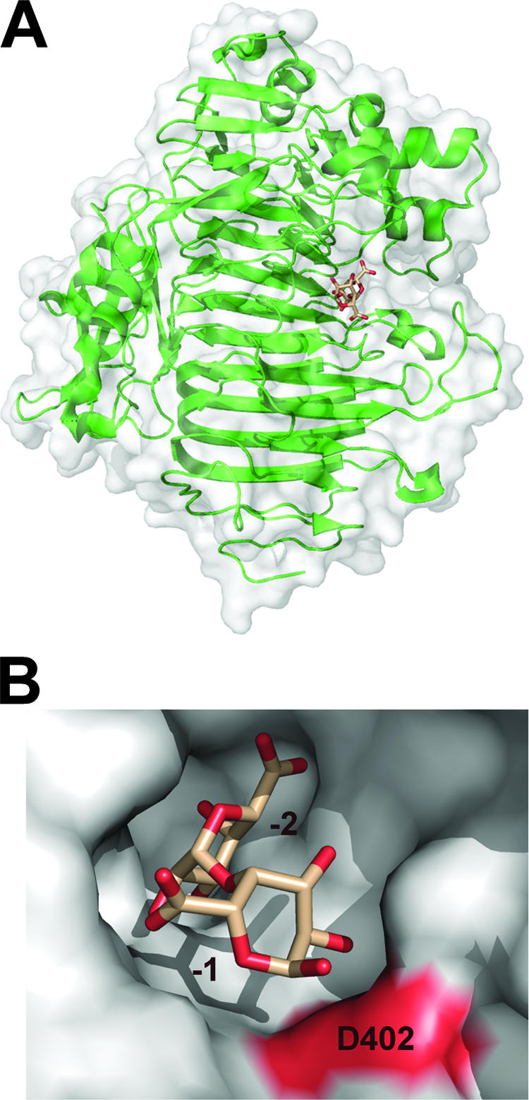
    1. Abbott, D. W., and A. B. Boraston. 2007. The structural basis for exopolygalacturonase activity in a family 28 glycoside hydrolase. J. Mol. Biol. 3681215-1222. - PubMed
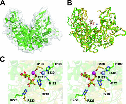
    1. Abbott, D. W., and A. B. Boraston. 2007. A family 2 pectate lyase displays a rare fold and transition metal-assisted beta-elimination. J. Biol. Chem. 28235328-35336. - PubMed
    1. Abbott, D. W., J. M. Eirin-Lopez, and A. B. Boraston. 2008. Insight into ligand diversity and novel biological roles for family 32 carbohydrate-binding modules. Mol. Biol. Evol. 25155-167. - PubMed
    1. Abbott, D. W., S. Hrynuik, and A. B. Boraston. 2007. Identification and characterization of a novel periplasmic polygalacturonic acid binding protein from Yersinia enterocolitica. J. Mol. Biol. 3671023-1033. - PubMed

LinkOut - more resources