Review
. 2008 Jun;72(2):317-64, table of contents.
doi: 10.1128/MMBR.00031-07.

Structure, function, and evolution of bacterial ATP-binding cassette systems

Affiliations
Review

Structure, function, and evolution of bacterial ATP-binding cassette systems

Amy L Davidson et al. Microbiol Mol Biol Rev. 2008 Jun.

Abstract

ATP-binding cassette (ABC) systems are universally distributed among living organisms and function in many different aspects of bacterial physiology. ABC transporters are best known for their role in the import of essential nutrients and the export of toxic molecules, but they can also mediate the transport of many other physiological substrates. In a classical transport reaction, two highly conserved ATP-binding domains or subunits couple the binding/hydrolysis of ATP to the translocation of particular substrates across the membrane, through interactions with membrane-spanning domains of the transporter. Variations on this basic theme involve soluble ABC ATP-binding proteins that couple ATP hydrolysis to nontransport processes, such as DNA repair and gene expression regulation. Insights into the structure, function, and mechanism of action of bacterial ABC proteins are reported, based on phylogenetic comparisons as well as classic biochemical and genetic approaches. The availability of an increasing number of high-resolution structures has provided a valuable framework for interpretation of recent studies, and realistic models have been proposed to explain how these fascinating molecular machines use complex dynamic processes to fulfill their numerous biological functions. These advances are also important for elucidating the mechanism of action of eukaryotic ABC proteins, because functional defects in many of them are responsible for severe human inherited diseases.


Figures

FIG. 1.
FIG. 1.
Conserved motifs in the ABC. Three characteristic motifs found in all ABC ATPases are represented by hatched red boxes. The Walker A motif and the Walker B motif form the nucleotide-binding fold of the P-loop ATPase family. The signature motif, also called the C loop, is unique to ABC proteins and also interacts with ATP. Other characteristic motifs, including the Q loop and the H loop (also called the switch region), contain just one highly conserved residue and are represented by hatched green boxes. These residues make contacts with the γ-phosphate of ATP. In the context of the ABC dimer, the D loop makes contacts with Walker motif A of the other monomer. Sequences between the Q loop and the signature constitute a helical domain, also referred to as a structurally diverse region (SDR), that contains residues important for the interaction of ABC proteins with their membrane partners.
FIG. 2.
FIG. 2.
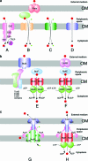
Schematic view of the organization of transport systems. In gram-negative bacteria, substrates can cross the outer membrane by facilitated diffusion through porins, which are trimeric channels. Red circles represent transported substrates, small green circles represent cotransported ions, and small blue circles represent phosphates. (a) Group translocators and secondary transporters. (A) PTSs. PTSs consist of a set of cytoplasmic energy-coupling proteins and various integral membrane permeases/sugar phosphotransferases, each specific for a different sugar. The E. coli mannitol permease consists of two cytoplasmic domains (EIIA and EIIB) involved in mannitol phosphorylation and an integral membrane domain (EIIC) which is sufficient to bind mannitol but which transports mannitol at a rate that is dependent on phosphorylation of the EIIA and EIIB domains. The two other components are common to all PTS systems. The soluble enzyme I (EI) autophosphorylates in the presence of Mg2+. The histidine protein (HPr) is the energy-coupling protein and delivers phosphoryl groups from EI to the sugar-specific transporters (EIIs). (B) TRAP transporters. A periplasmic BP, which is unrelated to an ABC BP at the sequence level but similar in secondary structure, functions in association with two membrane components, namely, a large TM subunit involved in the translocation process and a smaller membrane component of unknown function. The driving force for solute accumulation is an electrochemical ion gradient, not ATP hydrolysis. (C) Ion-driven MFS transporters. These transporters typically consist of a single cytoplasmic membrane protein with 12 TM segments that couples transport of small solutes to existing gradients of ions, such as protons or sodium ions. Symporters pump two or more types of solutes in the same direction simultaneously, using the electrochemical gradient of one of the solutes as the driving force. Antiporters (not shown) are driven in a similar way, except that the solutes are transported in opposite directions across the membrane. (D) Uniporters transport one type of solute and are driven directly by the substrate gradient. (b) ABC import systems. (E) Vitamin B12 importer. The vitamin B12 uptake system of E. coli includes a high-affinity OMR, BtuB, that translocates the substrate through the outer membrane in an energy-dependent step that requires an active TonB-ExbB-ExbD complex. Substrates are captured by the periplasmic BP BtuF in the periplasmic space and presented to a cytoplasmic complex made of two copies each of BtuC and BtuD. This complex mediates the ATP hydrolysis-dependent translocation of vitamin B12 into the cytoplasm. (F) Maltose-maltodextrin importer. The transport of maltodextrins larger than maltotriose through the outer membrane requires the trimeric maltoporin LamB. Substrates are captured by the maltose-BP MalE in the periplasmic space and presented to a cytoplasmic complex made of MalF, MalG, and two copies of MalK. (c) Comparison between secondary RND and primary ABC export systems. (G) AcrAB-TolC exporter. This hypothetical model of the RND family AcrA-AcrB-TolC drug efflux pump is based on the trimeric structures determined for TolC and AcrB. TolC is predicted to contact the apex of the AcrB trimer. Two molecules of the MFP AcrA are shown, but it is probable that this protein exists as higher-order oligomers in the complex. Hydrophobic drugs are probably pumped out of the membrane lipid bilayer coupled to the downhill movement of protons across the cytoplasmic membrane. (H) Hemolysin HlyBD-TolC exporter. This hypothetical assembled model consists of a TolC trimer, a dimer of the IM-ABC protein HlyB, and the MFP HlyD. The exact oligomeric state of HlyD is not known accurately, though it may be trimeric. The TM and ABC domains of HlyB are represented by red rectangles and green circles, respectively. Hemolysin is translocated through the envelope by an ATP hydrolysis-dependent process.
FIG. 3.
FIG. 3.
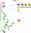
Simplified neighbor-joining tree of ABC domains. For clarity, only the main branches that point to ABC families are drawn. The major subdivisions of the tree correspond to the three classes of ABC systems, whose schematic structural representations are given in the right part of the figure. ABC domains are shown with green circles, and IM domains are shown with differently colored rectangles. For the sake of simplicity, accessory proteins (BPs, MFPs, and OMFs) are omitted. Class 1 (red branches) systems have fused ABC and IM domains, corresponding mainly to exporters. IM domains shown in purple represent ABC transporters with the IM domain at the N terminus, corresponding to IM-ABC and (IM-ABC)2 topologies. N- and C-terminal domains are symbolized by N and C, respectively. IM domains shown in orange represent ABC transporters with the IM domain at the C terminus, corresponding to ABC-IM and (ABC-IM)2 topologies. Class 1 contains two atypical families of systems, CCM and MCM, with a different structural organization. Class 2 (blue branches) systems have tandemly repeated ABC domains and no known TM domains (ABC2 topology), corresponding to proteins involved in nontransport processes. The UVR family was omitted during the generation of the tree because large domain insertions within the ABC domains prevent the establishment of the multiple sequence alignment. However, binary comparisons established the relationship between UVR proteins and class 2 systems. Class 3 (green branches) systems have IM (red rectangles) and ABC (green circles) domains carried by independent polypeptide chains, corresponding mainly to importers. For class 3, systems that could be exporters are shown in boxes. Family names are abbreviated according to the conventions used in Table 1 and throughout the text. See Table 1 for the abbreviations of family names and for functional descriptions. OPN-D, OPN-F, HAA-F, and HAA-G correspond to the two different ABC subunits of the OPN and HAA systems, respectively. MOS-N and MOS-C correspond to the N- and C-terminal ABC domains of MOS family ATPases. The scale at the top of the figure corresponds to 5% divergence per site between sequences.
FIG. 4.
FIG. 4.
Hypothetical scenario for the evolution of ABC systems. The figure uses the same topological representations and color coding as those used in Fig. 3. The hypothetical LUCA is predicted to possess all classes of ABC systems. The cell membrane is represented as a thick black line surrounding the intracellular medium (blue). The eukaryotic cell nucleus is a gray oval. The eukaryotic organelle is represented as a bacterium to symbolize its endosymbiotic origin. The arrows symbolize the evolutionary relationships between archaea, bacteria, and eukaryotes. The question mark under the arrow joining archaea and eukaryotes recalls the hypothesis that the ancestral eukaryotic cell arose by a unique endosymbiotic event involving engulfment of an archaebacterium by a gram-negative eubacterial host prior to the other endosymbiotic events leading to the appearance of organelles. Alternatively, eukaryotes may have arisen directly from the LUCA via an unidentified transient archaebacterium. A class 3 transporter, represented by a hatched pattern, recalls the hypothesis that these systems were acquired by eukaryotes and subsequently lost. See details of the scenario in the text.
FIG. 5.
FIG. 5.
Genomic distribution of ABC systems in living organisms. The plot shows the number of ABC ATPases versus the number of total genes in completely sequenced genomes. The number of ABC ATPases per genome (which roughly reflects the number of ABC systems) is plotted against the total number of genes (purple dots, archaea; blue dots, bacteria; green dots, eukaryotes). Selected genomes with exceptionally large or small numbers of ABC proteins are indicated with circles on the graph and are discussed in the text.
FIG. 6.
FIG. 6.
Structure of an ATP-bound ABC dimer. The structure of a MalK homodimer with two ATPs bound (PDB accession no. 1Q12) is shown. Each NBD consists of two subdomains, a RecA-like subdomain (green) and a helical subdomain (blue). A C-terminal RD (not present in all ABC proteins) is shown in yellow. Corresponding domains in the second MalK subunit follow the same color scheme but are rendered in lighter colors. Two ATPs, represented as a ball-and-stick model, are bound between the NBDs. The Walker A motif is shown in red.
FIG. 7.
FIG. 7.
Three structures of the MalK dimer. In the absence of nucleotide, the two NBDs of MalK are separated from each other, held as a dimer primarily through contacts between the C-terminal RDs. In the presence of ATP, the NBDs are closed, permitting ATP hydrolysis to occur. In the ADP-bound state, the NBDs are again separated, suggesting a possible cycle for hydrolysis-driven conformational change. Coloring is the same as that in Fig. 6. (Reprinted from reference with permission of the publisher. Copyright 2005 National Academy of Sciences, U.S.A.)
FIG. 8.
FIG. 8.
Structures of periplasmic maltose-BP. Structures of maltose-BP in the open, unliganded (blue; PDB accession no. 1ANF) and closed, maltose-bound (pink; PDB accession no. 1OMP) conformations are aligned based on the positions of α-carbons in the C lobe. The offset of the N lobes illustrates the domain rotation induced by ligand binding to class I and class II BPs.
FIG. 9.
FIG. 9.
Thermodynamic cycle for ligand binding to a periplasmic-BP. Munliganded and Mliganded are the equilibrium allosteric constants for the interconversion of the open and closed forms of the BP in the absence and presence of ligand (filled circles). Kopen and Kclosed are the theoretical equilibrium association constants for the binding of ligand to open and closed forms of the BP. (Reprinted from reference .)
FIG. 10.
FIG. 10.
Balancing interface in a BP. Cartoons of maltose-BP in open (unliganded) and closed (liganded) conformations depict the ligand-binding cleft and balancing interface. (Reprinted from reference with permission of the publisher.)
FIG. 11.
FIG. 11.
Concerted model for maltose transport. In the absence of maltose, the MalFGK2 transporter rests in a conformation in which the NBD dimer interface is open and the translocation pathway is exposed only to the cytoplasm (P-closed conformation). On binding of maltose, the periplasmic maltose-BP undergoes a conformational change from open to closed, and interaction with the closed maltose-BP in the presence of ATP triggers a global conformational change in which the NBDs close to promote ATP hydrolysis, maltose-BP becomes tightly bound to MalFGK2, and both maltose-BP and MalFGK2 open at the periplasmic surface of the membrane to facilitate the transfer of substrate from maltose-BP to a binding site in the membrane (P-open conformation). Following ATP hydrolysis, which destabilizes the NBD dimer, the transporter returns to the resting state and maltose completes its translocation across the membrane. (Adapted from reference with permission of the publisher. Copyright 2001 National Academy of Sciences, U.S.A.)
FIG. 12.
FIG. 12.
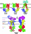
Structures of four importers. All transporters consist of four subunits, including a homodimer or heterodimer of IM subunits and a homodimer of NBD subunits. The ModB2C2 transporter is cocrystallized with ModA, a periplasmic BP in a closed, liganded conformation (cyan). The ligand, molybdate, is represented with a space-filling model. Maltose-BP (purple) is trapped in complex with MalFGK2 by using an E159Q substitution in MalK to prevent ATP hydrolysis (343). Nucleotide (ATP) is present only in MalFGK2E. A space-filling rendition of MalFGK2 is cut away to reveal an occluded pocket containing bound maltose. (Lower panel modified from reference .)
FIG. 13.
FIG. 13.
Cytochrome c-type biogenesis through the CcmA to CcmH and CydDC pathways. CcmA and CcmB (CcmA2B2) may form an ABC transporter for the transport of an as yet undiscovered molecule, and CcmC was proposed to export heme (represented as a cross with a dot) into the periplasm. Alternatively, a CcmA2BC complex may be involved in the export of heme. After transport, heme is transferred to the heme chaperone CcmE. CcmE is proposed to shuttle between CcmC and CcmF, and the protein CcmD has been shown to facilitate the interaction between CcmC and CcmE. Heme is transferred from CcmE to CcmF, which, in association with CcmG and CcmH, forms a heme-lyase complex. CcmG and CcmH maintain the apocytochrome (apo-cytc; green oval) in a reduced state. CcmG interacts with a periplasmic domain of the DsbD protein, which transfers electrons from the cytoplasm to the periplasm. Export of reductants such as cysteine or glutathione by the CydDC ABC transporter would help to maintain periplasmic redox homeostasis. This figure is based on data summarized in references and .
FIG. 14.
FIG. 14.
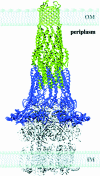
Model depicting export across two membranes. Both RND and ABC transporters interact with OMFs and MFPs to form a continuous translocation pathway across both the inner and outer membranes of gram-negative bacteria. This model offers one possible juxtaposition, using the structures of the OMF TolC trimer, the MFP MexA, and the RND AcrB trimer. (Reprinted from reference with permission of the publisher. Copyright 2004 National Academy of Sciences, U.S.A.)
FIG. 15.
FIG. 15.
Schematic representation of the drug-binding sites in P-glycoprotein, as proposed by Shapiro and Ling. Some drugs bind exclusively in the H site (blue) or in the R site (yellow), while others may be accommodated in both sites (red). This figure is based on data from reference .
FIG. 16.
FIG. 16.
Structure of an exporter. Sav1866 is a homodimer of two IM-ABC subunits. IM domain 1 (green) is fused to NBD1 (blue), and IM domain 2 (red) is fused to NBD2 (yellow). Bound ADP is represented with a ball-and-stick model. The front view (left) reveals a cleft in the IM region that would be exposed to the outer leaflet of the lipid bilayer. TM helices are predicted to extend well into the cytoplasm, creating an ICD. The side view (right) reveals the intertwisting of TM helices that places the coupling helix of one subunit into the cleft of the NBD of the second subunit in an example of domain swapping.
FIG. 17.
FIG. 17.
Undecaprenyl-linked O antigen polysaccharide precursors (O-PS), initiated by a WecA homologue and elongated at the nonreducing end (black dots), are polymerized in the cytosol by the Wbd glycosyltransferases, shown in purple. The ABC transporter formed by Wzm and Wzt is required for transfer of these undecaprenyl-linked polymers to the periplasmic face of the membrane (left side of the figure). The core-KDO-lipid A complex is synthesized by the Lpx proteins, and the addition of sugar residues (blue dots) that constitute the core polysaccharide is catalyzed by the Waa proteins. KDO-lipid A is flipped from the inner face to the outer face of the membrane by the MsbA ABC transporter (right side of the figure). The WaaL ligase, shown in green, ligates O-PS to KDO-lipid A to form LPS. Export of LPS to the outer membrane is dependent on the LptABC proteins (formerly called YhbN, YhbG, and YrbK), forming an ABC transporter with LptFG (formerly called YjgP and YjgQ) (middle part of the figure). Additional proteins (?) might participate in this step. The LptDE proteins (formerly called Imp and Rlp) are required to translocate LPS to the outer face of the outer membrane. This figure is based on data from references , , , and .

References

    1. Akama, H., M. Kanemaki, M. Yoshimura, T. Tsukihara, T. Kashiwagi, H. Yoneyama, S. Narita, A. Nakagawa, and T. Nakae. 2004. Crystal structure of the drug discharge outer membrane protein, OprM, of Pseudomonas aeruginosa: dual modes of membrane anchoring and occluded cavity end. J. Biol. Chem. 27952816-52819. - PubMed
    1. Akama, H., T. Matsuura, S. Kashiwagi, H. Yoneyama, S. Narita, T. Tsukihara, A. Nakagawa, and T. Nakae. 2004. Crystal structure of the membrane fusion protein, MexA, of the multidrug transporter in Pseudomonas aeruginosa. J. Biol. Chem. 27925939-25942. - PubMed
    1. Alaimo, C., I. Catrein, L. Morf, C. L. Marolda, N. Callewaert, M. A. Valvano, M. F. Feldman, and M. Aebi. 2006. Two distinct but interchangeable mechanisms for flipping of lipid-linked oligosaccharides. EMBO J. 25967-976. - PMC - PubMed
    1. Albers, S. V., S. M. Koning, W. N. Konings, and A. J. Driessen. 2004. Insights into ABC transport in archaea. J. Bioenerg. Biomembr. 365-15. - PubMed
    1. Alekshun, M. N., and S. B. Levy. 2007. Molecular mechanisms of antibacterial multidrug resistance. Cell 1281037-1050. - PubMed

Publication types

MeSH terms

Substances

LinkOut - more resources