

Enrichment of C-terminal fragments in TAR DNA-binding protein-43 cytoplasmic inclusions in brain but not in spinal cord of frontotemporal lobar degeneration and amyotrophic lateral sclerosis
- PMID: 18535185
- PMCID: PMC2438296
- DOI: 10.2353/ajpath.2008.080003
Enrichment of C-terminal fragments in TAR DNA-binding protein-43 cytoplasmic inclusions in brain but not in spinal cord of frontotemporal lobar degeneration and amyotrophic lateral sclerosis
Abstract
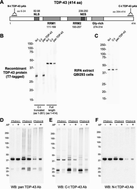
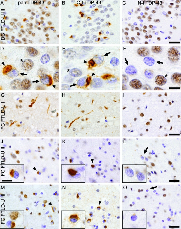
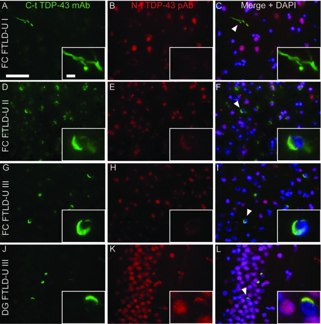
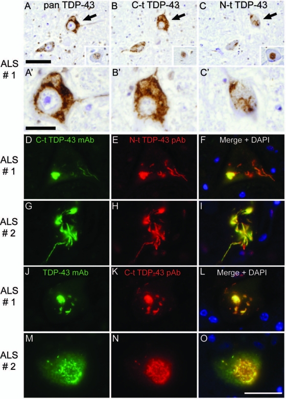
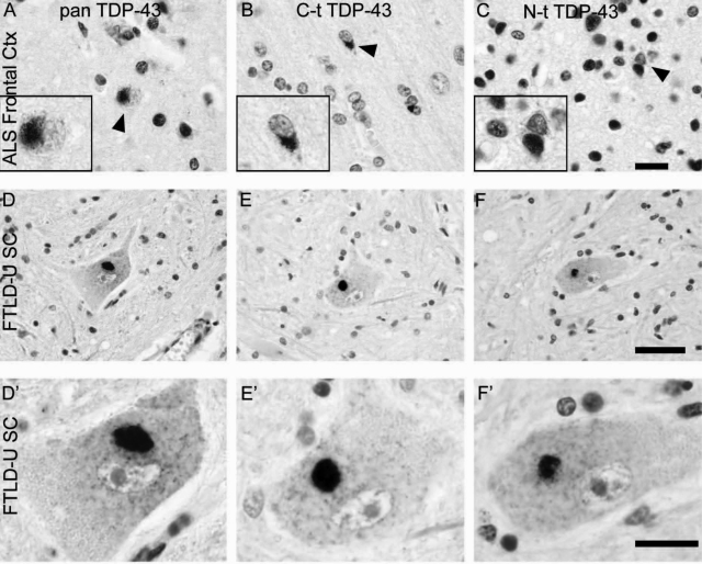
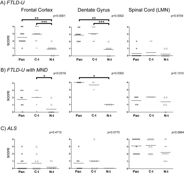
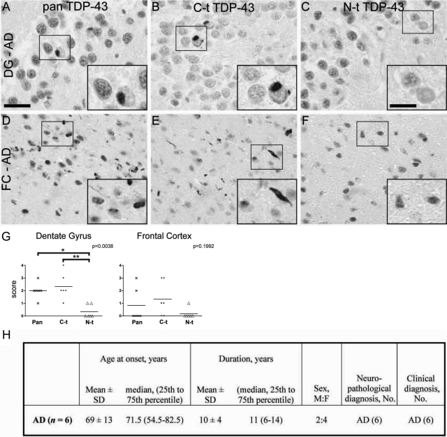
TAR DNA-binding protein (TDP-43) has been recently described as a major pathological protein in both frontotemporal dementia with ubiquitin-positive inclusions (FTLD-U) and amyotrophic lateral sclerosis. However, little is known about the relative abundance and distribution of different pathological TDP-43 species, which include hyperphosphorylated, ubiquitinated, and N-terminally cleaved TDP-43. Here, we developed novel N-terminal (N-t) and C-terminal (C-t)-specific TDP-43 antibodies and performed biochemical and immunohistochemical studies to analyze cortical, hippocampal, and spinal cord tissue from frontotemporal dementia with ubiquitin-positive inclusions and amyotrophic lateral sclerosis cases. C-t-specific TDP-43 antibodies revealed similar abundance, morphology, and distribution of dystrophic neurites and neuronal cytoplasmic inclusions in cortex and hippocampus compared with previously described pan-TDP-43 antibodies. By contrast, N-t-specific TDP-43 antibodies only detected a small subset of these lesions. Biochemical studies confirmed the presence of C-t TDP-43 fragments but not extreme N-t fragments. Surprisingly, immunohistochemical analysis of inclusions in spinal cord motor neurons in both diseases showed that they are N-t and C-t positive. TDP-43 inclusions in Alzheimer's disease brains also were examined, and similar enrichment in C-t TDP-43 fragments was observed in cortex and hippocampus. These results show that the composition of the inclusions in brain versus spinal cord tissues differ, with an increased representation of C-t TDP-43 fragments in cortical and hippocampal regions. Therefore, regionally different pathogenic processes may underlie the development of abnormal TDP-43 proteinopathies.
Figures
References
-
- Snowden JS, Neary D, Mann DM. Frontotemporal dementia. Br J Psychiatry. 2002;180:140–143. - PubMed
-
- Cairns NJ, Bigio EH, Mackenzie IR, Neumann M, Lee VMY, Hatanpaa KJ, White CL, III, Schneider JA, Grinberg LT, Halliday G, Duyckaerts C, Lowe JS, Holm IE, Tolnay M, Okamoto K, Yokoo H, Murayama S, Woulfe J, Munoz DG, Dickson DW, Ince PG, Trojanowski JQ, Mann DM. Neuropathologic diagnostic and nosologic criteria for frontotemporal lobar degeneration: consensus of the Consortium for Frontotemporal Lobar Degeneration. Acta Neuropathol. 2007;114:5–22. - PMC - PubMed
-
- McKhann GM, Albert MS, Grossman M, Miller B, Dickson D, Trojanowski JQ. Clinical and pathological diagnosis of frontotemporal dementia: report of the Work Group on Frontotemporal Dementia and Pick’s Disease. Arch Neurol. 2001;58:1803–1809. - PubMed
-
- Sampathu DM, Neumann M, Kwong LK, Chou TT, Micsenyi M, Truax A, Bruce J, Grossman M, Trojanowski JQ, Lee VMY. Pathological heterogeneity of frontotemporal lobar degeneration with ubiquitin-positive inclusions delineated by ubiquitin immunohistochemistry and novel monoclonal antibodies. Am J Pathol. 2006;169:1343–1352. - PMC - PubMed
-
- Neumann M, Mackenzie IR, Cairns NJ, Boyer PJ, Markesbery WR, Smith CD, Taylor JP, Kretzschmar HA, Kimonis VE, Forman MS. TDP-43 in the ubiquitin pathology of frontotemporal dementia with VCP gene mutations. J Neuropathol Exp Neurol. 2007;66:152–157. - PubMed
Publication types
MeSH terms
Substances
Grants and funding
LinkOut - more resources
Full Text Sources
Other Literature Sources
Medical
Research Materials
