Patches with links: a unified system for processing faces in the macaque temporal lobe
- PMID: 18535247
- PMCID: PMC8344042
- DOI: 10.1126/science.1157436
Patches with links: a unified system for processing faces in the macaque temporal lobe
Abstract
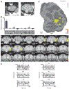
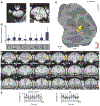
The brain processes objects through a series of regions along the ventral visual pathway, but the circuitry subserving the analysis of specific complex forms remains unknown. One complex form category, faces, selectively activates six patches of cortex in the macaque ventral pathway. To identify the connectivity of these face patches, we used electrical microstimulation combined with simultaneous functional magnetic resonance imaging. Stimulation of each of four targeted face patches produced strong activation, specifically within a subset of the other face patches. Stimulation outside the face patches produced an activation pattern that spared the face patches. These results suggest that the face patches form a strongly and specifically interconnected hierarchical network.
Figures


Similar articles
-
Effective Connectivity Reveals Largely Independent Parallel Networks of Face and Body Patches.Curr Biol. 2016 Dec 19;26(24):3269-3279. doi: 10.1016/j.cub.2016.09.059. Epub 2016 Nov 17. Curr Biol. 2016. PMID: 27866893
-
Functional compartmentalization and viewpoint generalization within the macaque face-processing system.Science. 2010 Nov 5;330(6005):845-51. doi: 10.1126/science.1194908. Science. 2010. PMID: 21051642 Free PMC article.
-
Parallel, multi-stage processing of colors, faces and shapes in macaque inferior temporal cortex.Nat Neurosci. 2013 Dec;16(12):1870-8. doi: 10.1038/nn.3555. Epub 2013 Oct 20. Nat Neurosci. 2013. PMID: 24141314 Free PMC article.
-
More Than the Face: Representations of Bodies in the Inferior Temporal Cortex.Annu Rev Vis Sci. 2022 Sep 15;8:383-405. doi: 10.1146/annurev-vision-100720-113429. Epub 2022 May 24. Annu Rev Vis Sci. 2022. PMID: 35610000 Review.
-
Rethinking cortical recycling in ventral temporal cortex.Trends Cogn Sci. 2024 Jan;28(1):8-17. doi: 10.1016/j.tics.2023.09.006. Epub 2023 Oct 17. Trends Cogn Sci. 2024. PMID: 37858388 Free PMC article. Review.
Cited by
-
Functional connectivity in category-selective brain networks after encoding predicts subsequent memory.Hippocampus. 2019 May;29(5):440-450. doi: 10.1002/hipo.23003. Epub 2018 Sep 2. Hippocampus. 2019. PMID: 30009477 Free PMC article.
-
The effects of electrical microstimulation on cortical signal propagation.Nat Neurosci. 2010 Oct;13(10):1283-91. doi: 10.1038/nn.2631. Epub 2010 Sep 5. Nat Neurosci. 2010. PMID: 20818384
-
Convolutional neural networks explain tuning properties of anterior, but not middle, face-processing areas in macaque inferotemporal cortex.Commun Biol. 2020 May 8;3(1):221. doi: 10.1038/s42003-020-0945-x. Commun Biol. 2020. PMID: 32385392 Free PMC article.
-
Neural representations of faces and body parts in macaque and human cortex: a comparative FMRI study.J Neurophysiol. 2009 May;101(5):2581-600. doi: 10.1152/jn.91198.2008. Epub 2009 Feb 18. J Neurophysiol. 2009. PMID: 19225169 Free PMC article.
-
The effect of face patch microstimulation on perception of faces and objects.Nat Neurosci. 2017 May;20(5):743-752. doi: 10.1038/nn.4527. Epub 2017 Mar 13. Nat Neurosci. 2017. PMID: 28288127 Free PMC article.
References
Publication types
MeSH terms
Grants and funding
LinkOut - more resources
Full Text Sources

