. 2008 Jul;118(7):2592-9.
doi: 10.1172/JCI33493.

Multipotential stem cells recapitulate human infantile hemangioma in immunodeficient mice

Affiliations

Multipotential stem cells recapitulate human infantile hemangioma in immunodeficient mice

Zia A Khan et al. J Clin Invest. 2008 Jul.

Abstract

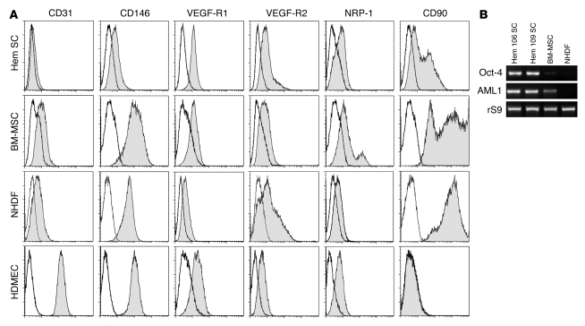
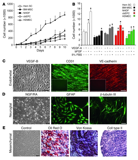
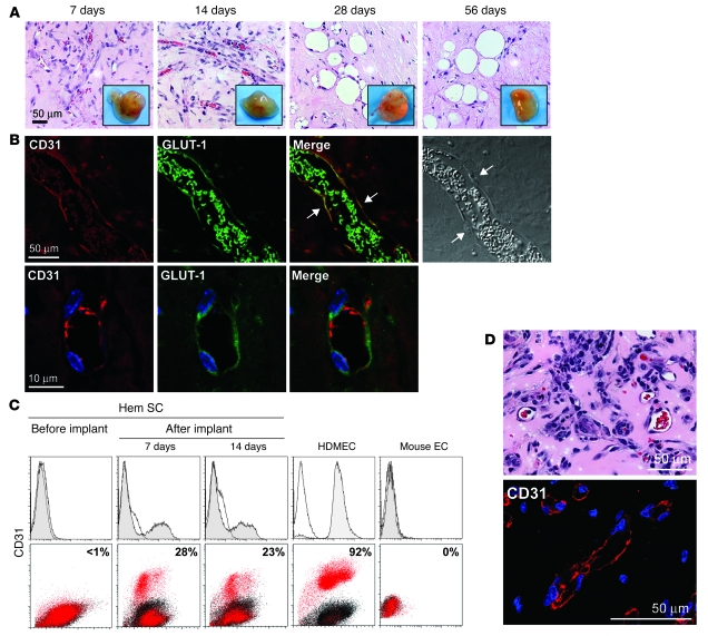
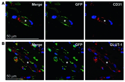
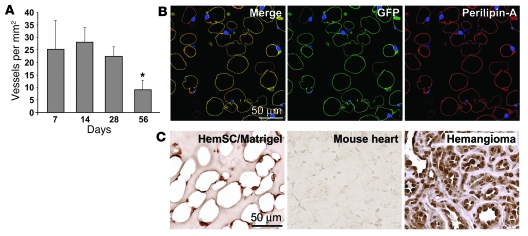
Infantile hemangioma is a benign endothelial tumor composed of disorganized blood vessels. It exhibits a unique life cycle of rapid postnatal growth followed by slow regression to a fibrofatty residuum. Here, we have reported the isolation of multipotential stem cells from hemangioma tissue that give rise to hemangioma-like lesions in immunodeficient mice. Cells were isolated based on expression of the stem cell marker CD133 and expanded from single cells as clonal populations. The CD133-selected cells generated human blood vessels 7 days after implantation in immunodeficient mice. Cell retrieval experiments showed the cells could again form vessels when transplanted into secondary recipients. The human vessels expressed GLUT-1 and merosin, immunodiagnostic markers for infantile hemangioma. Two months after implantation, the number of blood vessels diminished and human adipocytes became evident. Lentiviral expression of GFP was used to confirm that the hemangioma-derived cells formed the blood vessels and adipocytes in the immunodeficient mice. Thus, when transplanted into immunodeficient mice, hemangioma-derived cells recapitulated the unique evolution of infantile hemangioma--the formation of blood vessels followed by involution to fatty tissue. In summary, this study identifies a stem cell as the cellular origin of infantile hemangioma and describes for what we believe is the first time an animal model for this common tumor of infancy.


Figures

Figure 1
Figure 1. HemSCs express VEGF-Rs and mesenchymal cell marker CD90.
(A) Flow cytometric analysis of HemSCs, BM-MSCs, NHDFs, and HDMECs. Each cell type was grown in the EBM-2/20% FBS and assayed at passage 6. Gray histograms show cells labeled with FITC- or PE-conjugated antibodies. Black lines show isotype-matched control FITC- or PE-conjugated antibodies. Incubation with anti-CD31 and anti–VEGF-R2 was carried out following saponin permeabilization of HemSCs, BM-MSCs, and NHDFs. (B) RT-PCR analysis of Oct-4 and AML1 in 2 different HemSCs, Hem 106 and 109, with BM-MSCs and NHDFs shown for comparison. Ribosomal S9 (rS9) served as a control.
Figure 2
Figure 2. In vitro growth and multilineage differentiation of HemSCs.
(A) Proliferation of HemSCs in EBM-2/20% FBS over 10 days compared with normal endothelial and mesenchymal cells. (B) Proliferation in response to VEGF-A, bFGF, or 5% FBS for 24 hours in serum-free, growth factor–free EBM medium. *P < 0.05 compared with cells in serum-free, growth factor–free medium. (CE) Clonal HemSCs differentiated into endothelial (C), neuroglial (D), and mesenchymal (E) cells. Scale bars: 50 μm. Insets in C show CD31 and VE-cadherin immunostaining of cells induced in the absence of VEGF-B. All experiments were carried out with cells at passages 6–9.
Figure 3
Figure 3. HemSCs form CD31+GLUT-1+ blood vessels in vivo.
(A) Clonal HemSCs were suspended in Matrigel and injected s.c. into nude mice. H&E sections of explants at 7, 14, 28, and 56 days with insets showing explants at the corresponding time points. Scale bar: 50 μm. Passage 9 clonal HemSCs were used. (B) Immunofluorescent staining of day 7 explants. Human CD31 (red) is shown on the left, followed by GLUT-1 (green), a merged image, and a phase contrast image. In the merged image, arrows point to cells along the lumen of the blood vessel that are double-labeled for human CD31 and GLUT-1. rbc in the lumen are also positive for GLUT-1 (green). In the phase image, arrows point to endothelial nuclei. The bottom row shows higher magnification images. Scale bars: 50 μm (top row); 10 μm (bottom row). (C) Flow cytometry of cells reisolated from Matrigel explants after 7 and 14 days in vivo. Cells were labeled with anti-human CD31 without permeabilization to detect surface-localized CD31. HemSCs before implantation were negative for CD31 (left panels). HDMECs and mouse ECs are shown as positive and negative control cells. (D) Retrieved CD31+ cells formed blood vessels in secondary recipient mice. Matrigel implants were removed after 14 days, sectioned, stained with H&E (top panel), and immunostained with anti-human CD31 (bottom panel). Vessel density was 78 ± 35 rbc-filled lumens/mm2. Scale bars: 50 μM.
Figure 4
Figure 4. GFP-labeled HemSCs form hemangioma blood vessels in vivo.
(A) TurboGFP-expressing HemSCs were injected in Matrigel into mice, explanted at day 14, and localized by staining for TurboGFP (green), CD31 (red), and DAPI (blue). (B) Sections were also stained for TurboGFP (green), GLUT-1 (red), and DAPI (blue). Asterisks mark the lumen of the blood vessels in panels A and B. Scale bars: 50 μM.
Figure 5
Figure 5. HemSCs form adipocytes in vivo.
(A) Clonal HemSCs were suspended in Matrigel and injected s.c. into nude mice for 7, 14, 28, and 56 days, as shown in Figure 3A. Blood vessels, identified in H&E sections as luminal structures containing rbc, were quantified at the 4 time points. *P < 0.05 compared with other time points. (B) GFP-labeled HemSCs were implanted as described in Figure 4 and analyzed after 56 days for expression of GFP and the adipocyte marker perilipin A. Right panel shows the merged image, middle panel shows GFP (green), and right panel shows perilipin A (red); all panels were stained with DAPI. (C) Sections from the 28-day time point were immunostained with an anti-human nuclear antigen antibody using an HRP-conjugated secondary antibody and a diaminobenzidine substrate. Mouse heart tissue sections showed no immunostaining while nuclei in human hemangioma tissue were uniformly positive, confirming the specificity of anti-human nuclear antigen. Scale bars: 50 μM.

References

    1. Mulliken J.B., Fishman S.J., Burrows P.E. Vascular anomalies. Curr. Probl. Surg. 2000;37:517–584. doi: 10.1016/S0011-3840(00)80013-1. - DOI - PubMed
    1. Bischoff J. Monoclonal expansion of endothelial cells in hemangioma: an intrinsic defect with extrinsic consequences? Trends Cardiovasc. Med. 2002;12:220–224. doi: 10.1016/S1050-1738(02)00165-2. - DOI - PubMed
    1. Boye E., et al. Clonality and altered behavior of endothelial cells from hemangiomas. J. Clin. Invest. 2001;107:745–752. doi: 10.1172/JCI11432. - DOI - PMC - PubMed
    1. Bielenberg D.R., et al. Progressive growth of infantile cutaneous hemangiomas is directly correlated with hyperplasia and angiogenesis of adjacent epidermis and inversely correlated with expression of the endogenous angiogenesis inhibitor, IFN-beta. Int. J. Oncol. 1999;14:401–408. - PubMed
    1. Berard M., et al. Vascular endothelial growth factor confers a growth advantage in vitro and in vivo to stromal cells cultured from neonatal hemangiomas. Am. J. Pathol. 1997;150:1315–1326. - PMC - PubMed

Publication types

MeSH terms