. 2008 Jul 1;146A(13):1637-54.
doi: 10.1002/ajmg.a.32293.

Consistent chromosome abnormalities identify novel polymicrogyria loci in 1p36.3, 2p16.1-p23.1, 4q21.21-q22.1, 6q26-q27, and 21q2

Affiliations

Consistent chromosome abnormalities identify novel polymicrogyria loci in 1p36.3, 2p16.1-p23.1, 4q21.21-q22.1, 6q26-q27, and 21q2

William B Dobyns et al. Am J Med Genet A. .

Abstract

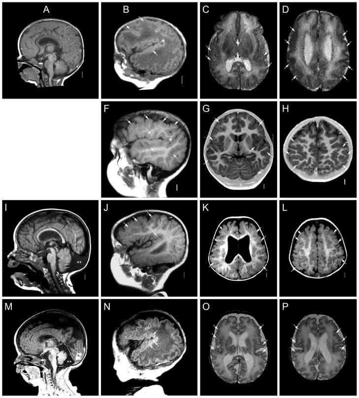
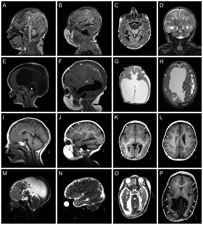
Polymicrogyria is a malformation of cortical development characterized by loss of the normal gyral pattern, which is replaced by many small and infolded gyri separated by shallow, partly fused sulci, and loss of middle cortical layers. The pathogenesis is unknown, yet emerging data supports the existence of several loci in the human genome. We report on the clinical and brain imaging features, and results of cytogenetic and molecular genetic studies in 29 patients with polymicrogyria associated with structural chromosome rearrangements. Our data map new polymicrogyria loci in chromosomes 1p36.3, 2p16.1-p23, 4q21.21-q22.1, 6q26-q27, and 21q21.3-q22.1, and possible loci in 1q44 and 18p as well. Most and possibly all of these loci demonstrate incomplete penetrance and variable expressivity. We anticipate that these data will serve as the basis for ongoing efforts to identify the causal genes located in these regions.


Figures

Figure 1
Figure 1
Brain images from four patients with deletion 1p36 including LP98-109 (A–D), LP99-072 (E–H), LP99-182 (I–L) and LR01-380 (M–P). These images demonstrate extensive areas of polymicrogyria (PMG, white or black arrows in many images) characterized by an irregular gyral pattern with abnormally wide sulci that are often too shallow or deep, as well as variably thick cortex. The PMG appears most severe in the posterior frontal and perisylvian regions and may be asymmetric. Here, the cortical malformation appears symmetric in two patients (C–D, O–P), mildly asymmetric in one (K–L) and strikingly asymmetric in another (G–H). In both asymmetric patients, the PMG is more severe on the right (left side of image as shown). Midline sagittal images show a mildly thin corpus callosum (arrowheads in A, E, I), and parasagittal images demonstrate an extended sylvian fissure (asterisks in B, F, N).
Figure 2
Figure 2
Brain images from patients with dup 2p16-p23 (A–D, LP99-112 and E–H, LR00-173), and del 4q21-q22 or q23 (I–L, LR04-022a2 and M–P, LR07-256). These images show extensive areas of PMG (white arrows in many images) that appear most severe in the posterior frontal and perisylvian regions, but are symmetric in all patients shown. Midline sagittal images show mild (I) or moderate (M) cerebellar vermis hypoplasia (CVH), as the bottom of the vermis (top white line in I and M) does not extend down to the level of the obex (bottom white line in I and M). The cisterna magna (double asterisks) is mildly prominent in one (M) and enlarged with an enlarged posterior fossa consistent with so-called mega-cisterna magna (I) in the other.
Figure 3
Figure 3
Brain images from four patients with deletion 6q26 including LR05-261 (A–D), LR00-218 (E–H), LP99-104a1 (I–L) and LP99-104a2 (M–P). These images show extensive areas of PMG (white arrows in many images) that is more difficult to appreciate than in other figures, but appears most severe in the temporal lobes and perisylvian regions. Midline sagittal images show thin and short corpus callosum (arrowheads in A and M) and CVH (arrowhead in E but also mildly small in A). Two patients had hydrocephalus (G and H, P).
Figure 4
Figure 4
Brain images from patients with del 18p (A–D, LP99-062), del 18p and 21q2 (E–H, LR00-219), and del 21q2 (I–L, LR05-078). These images show variable areas of PMG (white or black arrows in many images). In the girl with del 18p, the cortical abnormality involves the perisylvian and posterior regions on the right (C, D). In the boy with del 18p plus del 21q2, the PMG involves the entire cortex except for the mesial frontal lobes and appears most severe in the posterior frontal and perisylvian regions (G, H). In the girl with del 21q2, the PMG involves only the perisylvian regions (K, L). Midline sagittal images show dysplastic corpus callosum in one patient consisting of absent rostrum and small genu (arrowhead in I).

References

    1. Ahlbom BE, Sidenvall R, Anneren G. Deletion of chromosome 21 in a girl with congenital hypothyroidism and mild mental retardation. Am J Med Genet. 1996;64:501–505. - PubMed
    1. Alanay Y, Aktas D, Utine E, Talim B, Onderoglu L, Caglar M, Tuncbilek E. Is Dandy-Walker malformation associated with “distal 13q deletion syndrome”? Findings in a fetus supporting previous observations. Am J Med Genet Part A. 2005;136A:265–268. - PubMed
    1. Aligianis IA, Johnson CA, Gissen P, Chen D, Hampshire D, Hoffmann K, Maina EN, Morgan NV, Tee L, Morton J, et al. Mutations of the catalytic subunit of RAB3GAP cause Warburg Micro syndrome. Nat Genet. 2005;37:221–223. - PubMed
    1. Alkan M, Ramelli GP, Hirsiger H, Keser I, Remonda L, Buhler EM, Moser H. Presumptive monosomy 21 with neuronal migration disorder re-diagnosed as de novo unbalanced translocation t(18p;21q) by fluorescence in situ hybridisation. Genet Couns. 2002;13(2):151–6. - PubMed
    1. Amor DJ, Leventer RJ, Hayllar S, Bankier A. Polymicrogyria associated with scalp and limb defects: variant of Adams-Oliver syndrome [In Process Citation] Am J Med Genet. 2000;93:328–334. - PubMed

Publication types

MeSH terms