

Calcification of the internal elastic lamina of coronary arteries
- PMID: 18536656
- PMCID: PMC7029955
- DOI: 10.1038/modpathol.2008.89
Calcification of the internal elastic lamina of coronary arteries
Abstract
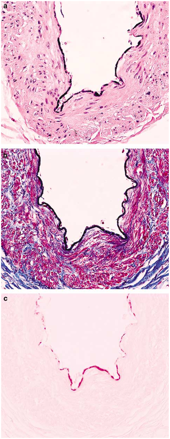
Two well-recognized patterns of calcification occur in large- and medium-sized arteries, intimal calcification associated with atherosclerosis and medial calcification described by Mönckeberg. Calcification limited to the internal elastic lamina is a third pattern of calcification not previously reported in coronary arteries. Here we describe 19 cases of coronary artery internal elastic lamina calcification. We serially sectioned and examined the coronary arteries of 66 patients with advanced AIDS and 27 HIV- controls with other chronic illnesses. We observed calcification of the internal elastic lamina in 10 HIV+ patients and 9 controls. HIV- patients with internal elastic lamina calcification were significantly older than HIV- patients without it (P=0.008) and HIV+ patients with it (P=0.006). Occasionally, calcification encroached on adjacent intimal or medial tissue with mild fibrosis. There was frequent disruption of the internal elastic lamina but no evidence of inflammation. Calcification was the dominant histologic feature in all cases. Von Kossa, Alizarin red, and trichrome/elastic stains confirmed these findings. Patients with internal elastic lamina calcification often had extensive medical histories but did not suffer from chronic renal failure or other conditions known to cause calcium dysregulation. We describe coronary internal elastic lamina calcification in HIV+ patients and older HIV- adults. The clinical significance of this finding is unknown. It could lead to arterial stiffening and increased pulse pressure and could be mistaken for intimal calcification on coronary imaging. Internal elastic lamina calcification may result from premature aging due to HIV disease and chronic illness or from metabolic disorders in HIV+ patients.
Conflict of interest statement
Disclosure/conflicts of interest
None.
Figures


References
-
- Simpson CL, Lindley S, Eisenberg C, et al. Toward cell therapy for vascular calcification: osteoclast-mediated demineralization of calcified elastin. Cardiovasc Pathol 2007;16:29–37. - PubMed
-
- Chen NX, Moe SM. Uremic vascular calcification. J Investig Med 2006;54:380–384. - PubMed
-
- Floege J, Ketteler M. Vascular calcification in patients with end-stage renal disease. Nephrol Dial Transplant 2004;19(Suppl 5):V59–V66. - PubMed
-
- Mönckeberg JG. Ueber die reine Mediaverkalkung der Extremitaetenarterien und ihr Verhalten zur Arteriosklerose. Virchows Arch Pathol Anat 1903;171:141–167.
Publication types
MeSH terms
Substances
Grants and funding
LinkOut - more resources
Full Text Sources
Medical
