. 2008 Jul;40(7):921-6.
doi: 10.1038/ng.173. Epub 2008 Jun 8.

Ras-MAPK signaling promotes trophectoderm formation from embryonic stem cells and mouse embryos

Affiliations

Ras-MAPK signaling promotes trophectoderm formation from embryonic stem cells and mouse embryos

Chi-Wei Lu et al. Nat Genet. 2008 Jul.

Abstract

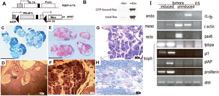
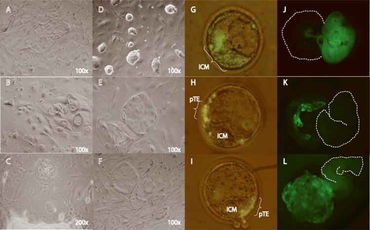
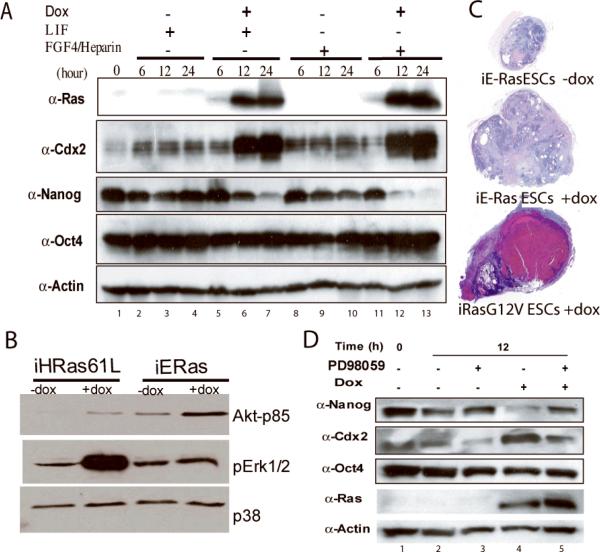
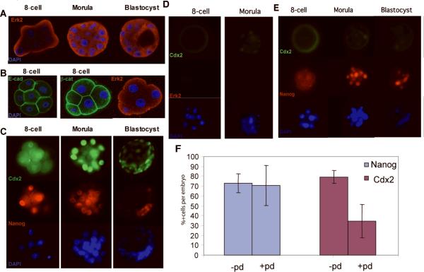
In blastocyst chimeras, embryonic stem (ES) cells contribute to embryonic tissues but not extraembryonic trophectoderm. Conditional activation of HRas1(Q61L) in ES cells in vitro induces the trophectoderm marker Cdx2 and enables derivation of trophoblast stem (TS) cell lines that, when injected into blastocysts, chimerize placental tissues. Erk2, the downstream effector of Ras-mitogen-activated protein kinase (MAPK) signaling, is asymmetrically expressed in the apical membranes of the 8-cell-stage embryo just before morula compaction. Inhibition of MAPK signaling in cultured mouse embryos compromises Cdx2 expression, delays blastocyst development and reduces trophectoderm outgrowth from embryo explants. These data show that ectopic Ras activation can divert ES cells toward extraembryonic trophoblastic fates and implicate Ras-MAPK signaling in promoting trophectoderm formation from mouse embryos.


Figures

Figure 1
Figure 1. Induction of expression of activated Ras promotes formation of trophoblastic tumors from ES cells
(A) Schema for generating inducible HRasQ61L ES cells. Ainv15 mouse embryonic stem cells have the rtTA integrated at the ROSA 26 locus on chromosome 6. HRasQ61L was inserted by Cre-mediated recombination of a targeting vector (plox) into the region of the HPRT locus so that it is expressed from the tetracycline response element (tetOP). Successful recombination regenerates an ATG-truncated neomycin (G418) resistance gene (Neo) driven by the pgk promoter (PGK-ATG). (B) Ras activation assay (Upstate Bioscience). Upper blot: Co-precipitation with Raf indicates expression of active GTP-bound Ras. Lower blot: Expression of total Ras can be detected by immunoblot in doxycycline-induced cells with anti-Ras antibody. Lane 1—Un-induced (no doxycycline); lane 2—induced with doxycycline. (C-H) tumors obtained from iRasES cells implanted in Rag2-/-γc-/- mice. Scale bars in C&E: 1cm. Magnification is indicated in each panel. (C) Cross-section of teratoma from control mice (no doxycyline); (D) Histology of teratoma in C showing tissue complexity (Hematoxylin and Eosin Staining); (E) Hemorrhagic tumors isolated from mice fed doxycycline; (F, G) Histology of tumors in E, showing clusters of giant cells (H&E staining). (H) Periodic Acid Schiff (PAS) staining of sections of tumors from doxycycline-induced animals, showing glycogen-rich granules. (I) RT-PCR analysis of gene expression of two teratomas from control animals (un-induced) and hemorrhagic tumors from two mice fed doxycycline (induced). Dihydrofolate reductase (DHFR) is used as a loading control for this analysis. α-fp(a-feto protein), c-actin (cardiac-actin) and pax6 are markers of differentiation toward endoderm, mesoderm and ectoderm, respectively. Markers for trophectoderm: for trophoblastic giant cells, pl-1 (placenta lactogen 1) and plAP (placenta alkaline phosphatase); for spongiotrophoblasts, tpbpα (trophoblastic specific protein alpha) and proliferin.
Figure 2
Figure 2. Trophoblastic stem cell establishment from iRasES cells
A-E: in vitro culture of iRas ESCs in various conditions: (A) culture in doxycycline (dox) and leukemia inhibitory factor (LIF) for 5 days. (B and C) Formation of giant cells (B) and syncytium (C) in iRasES culture when dox was present, without Fgf4 and LIF. (D) Colony morphology of iRas ES cells cultured in ES media supplemented with Fgf4 (without dox). (E) Colony morphology of iRasES cultured in Fgf4 and dox for two weeks. (F) Colonies from blastocyst-derived trophoblastic stem cells . G-L, iRasES cell derived trophoblastic stem cells (ES-TS) chimerize the trophectoderm and placenta of the developing embryo. (G) Parental iRasES cells labeled with the lipophilic dye pKH26 (Sigma), injected into 4-cell embryos, and examined by fluorescence microscopy at the blastocyst stage. Note green cells within ICM (representative image from 8 out of 8 injected embryos). (H) ES-TS cells derived from culture of iRasES cells in Fgf4 and dox, labeled with pKH26, injected into 4-cell embryos, and examined at the blastocyst stage. Note green cells in polar trophectoderm. (representative image from 5 out of 8 embryos injected. Green cells were undetectable in the other 3 embryos) (I) Blastocyst-derived TS cells labeled with pKH26, injected into 4-cell embryos, and examined at the blastocyst stage. (representative image from 3 out of 3 embryos injected) (J-L) Fluorescent images of embryos resulting from blastocysts chimerized by iRasES cells and iRasES cell-derived ES-TS cells. Cells were transduced by a lentivirus carrying Green Fluorescent Protein (GFP). (J) Chimeric embryo injected with parental iRasES cells. (K, L) Chimeric placental tissues of embryos injected with iRasES cell-derived ES-TS cells. Margins of embryo or dissected placental tissues are outlined.
Figure 3
Figure 3. Expression of Cdx2, Nanog, and Oct4 in response to Ras/MapK signaling in ES cells
(A) iRasES cells cultured as indicated for 6, 12 and 24 hours. Cell lysates were analyzed by immunoblot to detect protein levels of Ras, Cdx2, Nanog, Oct4 and Actin. (B) Cell lysates from iRasES cells (H-RasQ61L) and iE-Ras ES cells prepared after 48 hours of culture, and immunoblotted with antibody to activated phospho-proteins, as indicated. (C) Tumors formed by ES cells with or without doxycycline-induction of ERas or H-RasG12V, as indicated. (D) Cell lysates of iRasES cells treated for 12 hours with doxycycline and/or the MapK inhibitor PD98095, analyzed by immunoblot with the antibodies indicated.
Figure 4
Figure 4. Ras-MapK signaling regulates Cdx2 expression and trophoblast outgrowth in mouse embryos
(A-E) Immunofluorescence images of mouse embryos at 8-cell, morula and blastocyst stage. (A) confocal image of embryos stained with antibodies against Erk2, showing apical distribution at 8 cell stage and diffuse staining of morula and blastocyst. (B) Distribution of E-cadherin and β-catenin in 8-cell stage embryo. Embryos were stained with primary antibodies against E-cadherin and β-catenin. The left panel shows an 8-cell stage embryo stained with E-cadherin alone. The right panel shows another embryo co-stained with β-catenin and Erk-2. Co-staining of E-cadherin and pErk2 is not shown due to cross-reactivity of the antibodies. (C) Standard epifluorescence images of embryos stained with anti-Cdx2 and anti-Nanog, counter-stained with Dapi-antifad gold. (D and E) Expression of Cdx2, Nanog and Erk2 in embryos exposed to the MapK inhibitor PD98095 (20μM) for 24 hours. (F) Percentage of Nanog and Cdx2 positive cells within morula-stage embryos with or without exposure to PD98095. 6 embryos were scored in non-treated experiments, 7 embryos were scored in PD98059 treated experiments. Total cell numbers and Nanog/Cdx2 positive cell numbers in two experiments are listed in supplemental table 1. Error bars reflect standard deviations. p=0.780917 for Nanog expression; p= 0.000201 for Cdx2 expression, by two-tailed Student test.

References

    1. Nagy A, et al. Embryonic stem cells alone are able to support fetal development in the mouse. Development. 1990;110:815–21. - PubMed
    1. Tanaka S, Kunath T, Hadjantonakis AK, Nagy A, Rossant J. Promotion of trophoblast stem cell proliferation by FGF4. Science. 1998;282:2072–5. - PubMed
    1. Kunath T, et al. Imprinted X-inactivation in extra-embryonic endoderm cell lines from mouse blastocysts. Development. 2005;132:1649–61. - PubMed
    1. Land H, Parada LF, Weinberg RA. Tumorigenic conversion of primary embryo fibroblasts requires at least two cooperating oncogenes. Nature. 1983;304:596–602. - PubMed
    1. Hahn WC, et al. Creation of human tumour cells with defined genetic elements. Nature. 1999;400:464–8. - PubMed

Publication types

MeSH terms