. 2008 Jul;40(7):862-70.
doi: 10.1038/ng.157. Epub 2008 Jun 8.

A Gata6-Wnt pathway required for epithelial stem cell development and airway regeneration

Affiliations

A Gata6-Wnt pathway required for epithelial stem cell development and airway regeneration

Yuzhen Zhang et al. Nat Genet. 2008 Jul.

Abstract

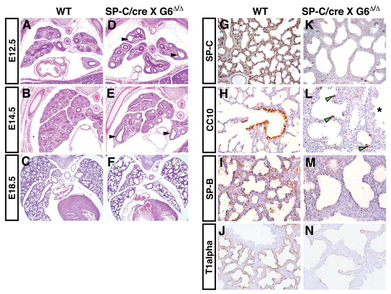
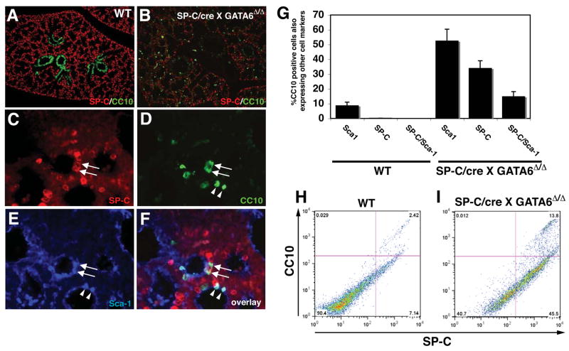
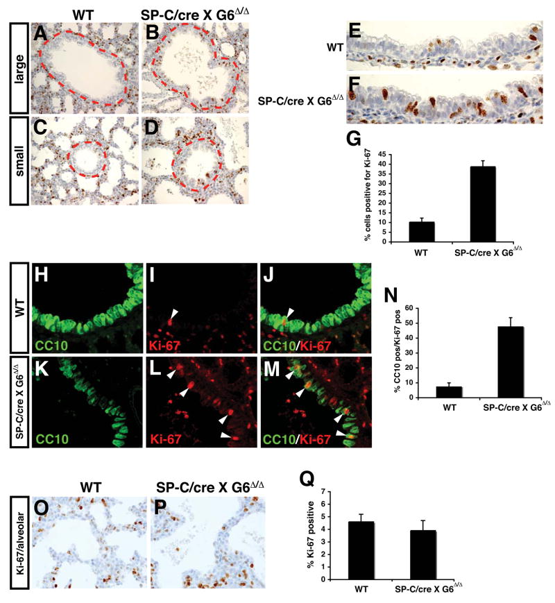
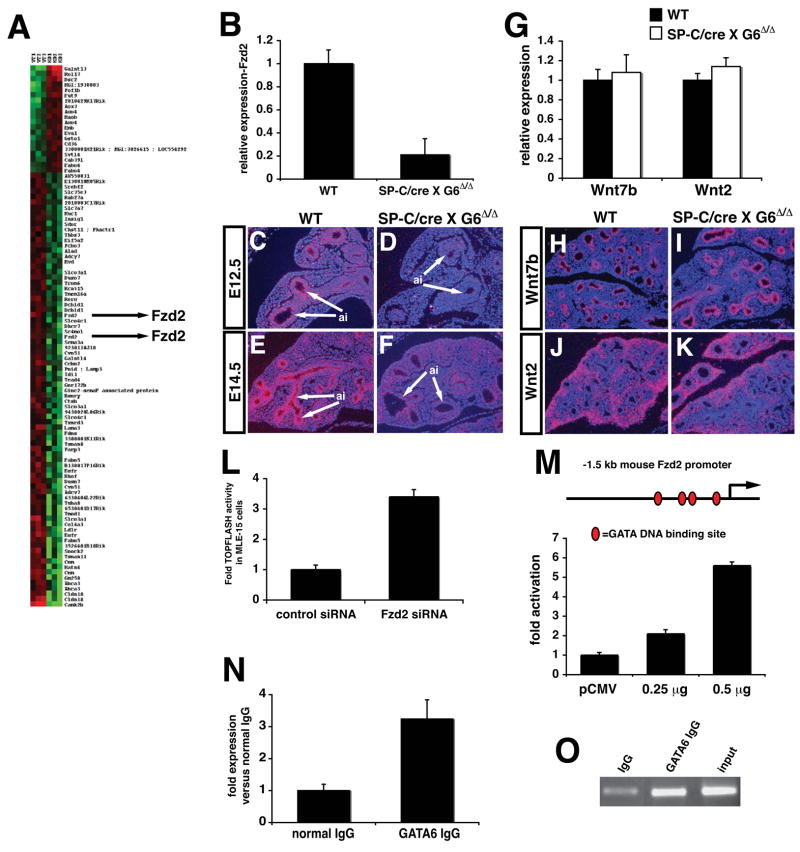
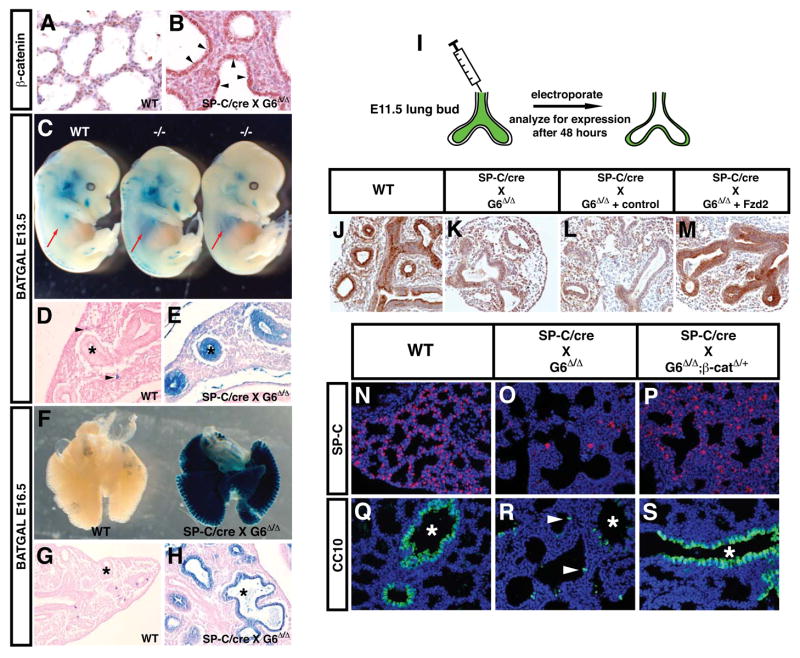
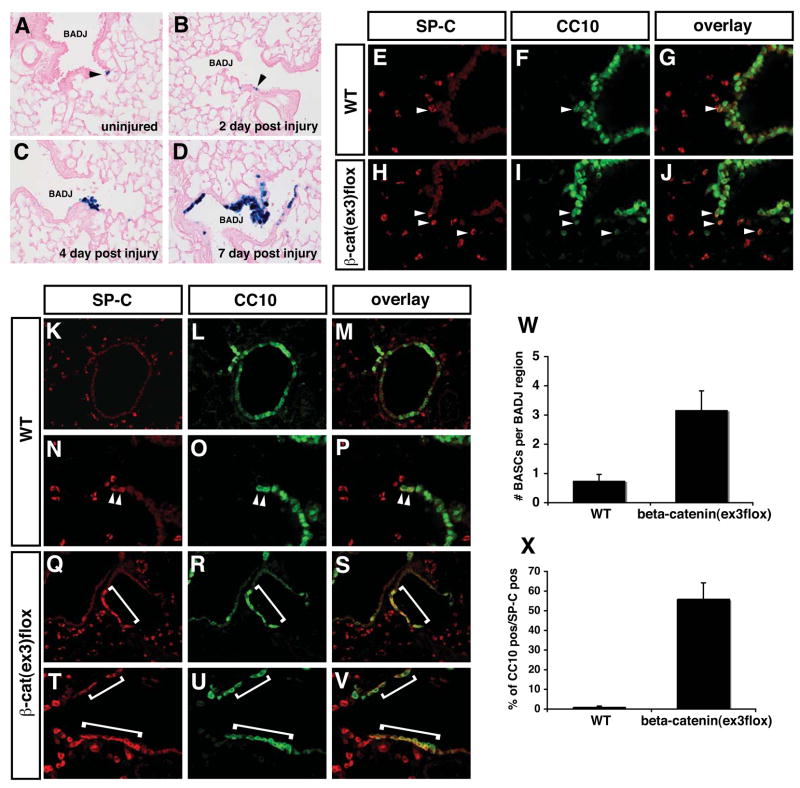
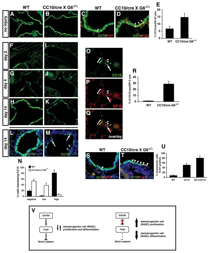
Epithelial organs, including the lung, are known to possess regenerative abilities through activation of endogenous stem cell populations, but the molecular pathways regulating stem cell expansion and regeneration are not well understood. Here we show that Gata6 regulates the temporal appearance and number of bronchioalveolar stem cells (BASCs) in the lung, its absence in Gata6-null lung epithelium leading to the precocious appearance of BASCs and concurrent loss in epithelial differentiation. This expansion of BASCs was the result of a pronounced increase in canonical Wnt signaling in lung epithelium upon loss of Gata6. Expression of the noncanonical Wnt receptor Fzd2 was downregulated in Gata6 mutants and increased Fzd2 or decreased beta-catenin expression rescued, in part, the lung epithelial defects in Gata6 mutants. During lung epithelial regeneration, canonical Wnt signaling was activated in the niche containing BASCs and forced activation of Wnt signaling led to a large increase in BASC numbers. Moreover, Gata6 was required for proper lung epithelial regeneration, and postnatal loss of Gata6 led to increased BASC expansion and decreased differentiation. Together, these data demonstrate that Gata6-regulated Wnt signaling controls the balance between progenitor expansion and epithelial differentiation required for both lung development and regeneration.


Figures

Figure 1
Figure 1. Loss of Gata6 leads to lung epithelial differentiation defects
Wild-type (WT) and Gata6 Δ/Δ :SP-C/cre mutants (SP-C/cre X G6 Δ/Δ) were examined by H+E staining (A–F) or by immunohistochemical staining for lung epithelial marker proteins.(G–N). Gata6 Δ/Δ :SP-C/cre mutants displayed dilated airways beginning as early as E12.5 (D and E, arrowheads). Gata6 Δ/Δ :SP-C/cre mutants showed decreased or absent expression of SP-C (G and K), CC10 (H and L), SP-B (I and M), and T lalpha (J and N). However, ectopic expression of the Clara cell marker protein CC10 was observed in distal regions of the airways of Gata6 Δ/Δ :SP-C/cre mutants (L, arrowheads). Asterisk indicates a large bronchiolar airway, which has reduced levels of CC10 expression in the mutants.
Figure 2
Figure 2. Loss of Gata6 in lung epithelium results in the precocious appearance of BASCs
Expression of SP-C and CC10 was analyzed at E18.5 in wild-type (A) and Gata6 Δ/Δ :SP-C/cre mutants (B–F) using double and triple immunofluorescent staining. Gata6 Δ/Δ :SP-C/cre mutants contained a large number of SP-C/CC10 double positive BASCs in airways (C and D, arrows and G) while wild-type lungs did not (A and G). Many of these double positive cells also expressed Sca1 (E and F, arrows). In addition to the SP-C/CCl0/Scal triple positive population, a CCl0/Scal population was also up-regulated in Gata6 Δ/Δ :SP-C/cre mutants (D–F, arrowheads, and G). FACS analysis was performed using SP-C and CC10 antibodies on both wild-type (H) and Gata6 Δ/Δ :SP-C/cre mutant (I) permeabilized lung epithelial cells at E18.5 and showed a significant increase in BASC numbers in the mutant lungs (2.4% versus 13.8%).
Figure 3
Figure 3. Increased proliferation in bronchiolar epithelium of Gata6 Δ/Δ :SP-C/cre mutants
Increased staining for Ki-67 was observed in Gata6 Δ/Δ :SP-C/cre mutants versus wild-type lungs in bronchiolar epithelium of both medium and small caliber airways (A–D, inside dotted line). This corresponded to a more disorganized appearance in this epithelium with multiple layers of cells growing on top of each other (E and F). Quantitation revealed an approximate four-fold increase in proliferation in bronchiolar epithelium (G). Double immunofluorescent microscopy showed that most of the increased cell proliferation was observed in cells weakly positive for CC10 in Gata6 Δ/Δ :SP-C/cre mutants (H–M, arrowheads, and N; green-CC10, red-Ki-67). Of note, CC10 expression as detected by immunofluorescent microscopy in these studies was very low and the data represent a longer exposure than used in wild-type samples. No increase in distal airway epithelial proliferation was observed in Gata6 Δ/Δ :SP-C/cre mutants as detected by Ki-67 immunostaining (O–Q).
Figure 4
Figure 4. Fzd2 is a target of Gata6 in lung epithelium and negatively regulates canonical Wnt signaling
Microarray studies were performed to compare gene expression profiles in wild-type versus Gata6 Δ/Δ :SP-C/cre mutants. A heat map representing a portion of the genes up or down regulated in Gata6 Δ/Δ :SP-C/cre mutants, highlighting Fzd2, is shown in (A). Q-PCR shows that Fzd2 expression is decreased approximately five-fold in Gata6 Δ/Δ :SP-C/cre mutants (B). In situ hybridization shows that Fzd2 expression is lost in airway epithelium but is still observed in mesenchyme of the lung in Gata6 Δ/Δ :SP-C/cre mutants (C–F, arrows). Q-PCR shows that Wnt7b and Wnt2 expression remains unchanged in Gata6 Δ/Δ :SP-C/cre mutants (G) while in situ hybridization shows that spatial expression of these Wnt ligands also remains unchanged (H–K). Knock-down of Fzd2 expression by siRNA in MLE-15 cells results in increased TOPFLASH activity (L). The proximal −1.5 kb Fzd2 promoter is trans-activated by Gata6 in a dose-dependent manner in NIH-3T3 cells (M). ChIP assays show that Gata6 is associated with the mouse Fzd2 promoter using Q-PCR (N) and as shown by agarose gel electrophoresis (O). ai-airways.
Figure 5
Figure 5. Increased canonical Wnt signaling in lung epithelium upon loss of Gata6 expression and rescue of these defects by re-expression of Fzd2 or decreased β-catenin expression
Activated (β-catenin protein expression is increased in Gata6 Δ/Δ :SP-C/cre mutant airway epithelium (A and B, arrows). The BAT-GAL lacZ Wnt reporter mice show increased canonical Wnt activity in the lungs of Gata6 Δ/Δ :SP–C/cre mutants at both E13.5 (C-E) and E16.5 (F–H). This activity is dramatic and can be observed through the chest wall of the mutants at E13.5 (C, arrows). This increase in Wnt signaling is confined to airway epithelium (E and H, asterisks). Residual lacZ expression is observed in wild-type littermates in scattered mesenchymal or epithelial cells (D and G, arrowheads). The airway lumen of E11.5 lung buds from both wild-type and Gata6 Δ/Δ :SP-C/cre mutants was injected with either control plasmid or an expression plasmid encoding mouse Fzd2 and then electroporated as described in Materials and Methods (I). SP-C expression was used as a indication of rescue of epithelial defects in Gata6 Δ/Δ :SP-C/cre mutants. As expected, SP-C expression was observed in the airway epithelium of the wild-type explants (J). Gata6 Δ/Δ:SP-C/cre mutants lacked SP-C expression (K). Electroporation of the control vector did not result in increased SP-C expression (L) whereas the Fzd2 expression plasmid resulted in re-expression of SP-C (M). Gata6 Δ/Δ :SP-C/cre mutants were crossed into the β-catenirflox/+ background and expression of SP-C and CC10 was analyzed in wild-type (N and Q), Gata6 Δ/Δ :SP-C/cre mutants (O and R), and Gata6 Δ/Δ-catenirflox/+:SP-C/cre mutants (P and S). As expected, Gata6 Δ/Δ:SP-C/cre mutants have significantly decreased SP-C and CC10 expression with ectopic CC10 expression in distal airways (O and R, arrowheads). However, loss of one allele of β-catenin leads to a marked increase in SP-C and CC10 expression in Gata6 Δ/Δ:SP-C/cre mutants and a decrease in ectopic CC10 expression (P and S). Asterisks-airway epithelium.
Figure 6
Figure 6. Canonical Wnt signaling is activated upon lung regeneration and forced activation of Wnt/β-catenin signaling leads to expansion of BASCs
BAT-GAL mice were subjected to naphthalene based lung airway injury to induce airway regeneration. In uninjured mice, only rare lacZ positive cells were observed in the BADJ region of the airways (A, arrowhead). After naphthalene injury and during the epithelial regeneration process, a dramatic increase in lacZ staining indicating increased canonical Wnt signaling is observed in cells within the BADJ niche were BASCs reside (B–D). SP-C/CC10 co-staining shows that BASC numbers are increased in the BADJ region of β-cateninΔex3:CC10/cre mice (E–J, arrows and W). Seven days after naphthalene induced lung injury and regeneration, a significant increase in BASC number is observed in β-cateninΔex3:CC10/cre mice (K–V, compare arrowheads to brackets). Quantitation reveals that approximately 50% of the cells regenerating in the bronchiolar airways are SP-C/CC10 double positive BASCs (X).
Figure 7
Figure 7. Gata6 regulates BASC expansion and differentiation in airway epithelial regeneration
Both wild-type and adult Gata6 Δ/Δ :CC10/cre mutants show normal expression of CC10 (A and B). However, Gata6 Δ/Δ :CC10/cre mutants showed increased proliferation (C–E). Napthalene injury of Gata6 Δ/Δ :CC10/cre mutants resulted in decreased Clara cell regeneration by 10 days after injury (F–K). CC10 positive cells that did regenerate were divided into two categories: low expressing cells and high expressing cells (L–N). Few if any CC10 high expressing cells regenerated in Gata6 Δ/Δ:CC10/cre mutants (N). This loss in Clara cell regeneration was accompanied by an increase in BASCs (O–R) and increased proliferation in the CC10 expressing cells that did regenerate (S–U). Gata6 regulates Fzd2 expression which in turn antagonizes canonical Wnt signaling in lung epithelia and allows proper epithelial differentiation and development of BASCs which is required for lung regeneration (V).

Comment in

  • Lung stem cells in the balance.
    Bellusci S. Bellusci S. Nat Genet. 2008 Jul;40(7):822-4. doi: 10.1038/ng0708-822. Nat Genet. 2008. PMID: 18583977 No abstract available.

References

    1. Cardoso WV. Molecular regulation of lung development. Annu Rev Physiol. 2001;63:471–94. - PubMed
    1. Cardoso WV, Lu J. Regulation of early lung morphogenesis: questions, facts and controversies. Development. 2006;133:1611–24. - PubMed
    1. Minoo P, Su G, Drum H, Bringas P, Kimura S. Defects in tracheoesophageal and lung morphogenesis in Nkx2.1 (−/−) mouse embryos. Dev. 1999:60–71. 5/0/209. - PubMed
    1. Wan H, et al. Compensatory roles of Foxa1 and Foxa2 during lungmorphogenesis. J Biol Chem. 2005;280:13809–16. - PubMed
    1. Yang H, Lu MM, Zhang L, Whitsett JA, Morrisey EE. GATA6 regulates differentiation of distal lung epithelium. Development. 2002;129:2233–46. - PubMed

Publication types

MeSH terms

Associated data

LinkOut - more resources