

doi: 10.1038/nmeth.1220.
Epub 2008 Jun 8.
Lifeact: a versatile marker to visualize F-actin
Affiliations
- PMID: 18536722
- PMCID: PMC2814344
- DOI: 10.1038/nmeth.1220
Item in Clipboard
Lifeact: a versatile marker to visualize F-actin
Nat Methods.
2008 Jul.
Abstract
Live imaging of the actin cytoskeleton is crucial for the study of many fundamental biological processes, but current approaches to visualize actin have several limitations. Here we describe Lifeact, a 17-amino-acid peptide, which stained filamentous actin (F-actin) structures in eukaryotic cells and tissues. Lifeact did not interfere with actin dynamics in vitro and in vivo and in its chemically modified peptide form allowed visualization of actin dynamics in nontransfectable cells.
Conflict of interest statement
Figures
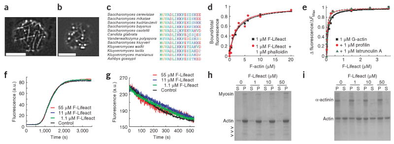
Identification and biochemical characterization of Lifeact. (a) TIRF microscopy image of Abp140-GFP in an unpolarized yeast cell. (b) Epifluorescence image of Lifeact-GFP in a yeast cell. Scale bars, 5 μm. (c) Alignment of the actin-binding sequence in fungi. (d) Bound/total fluorescence of F-Lifeact co-sedimented with rabbit muscle F-actin. (e) Fluorescence of pyrene-labeled G-actin in the presence of F-Lifeact. Fluorescence was normalized to maximum values. (f) Polymerization of 20% pyrene-labeled actin in the absence (control) or presence of indicated concentrations of F-Lifeact. (g) Depolymerization of 100% pyrene-labeled F-actin after dilution below 200 nM with F-Lifeact (control without Lifeact). (h,i) SDS PAGE of pellet (P) and supernatant (S) fractions of F-actin sedimented with myosin (h) and α-actinin (i) in the absence and presence of F-Lifeact. Arrowheads, myosin light chains.
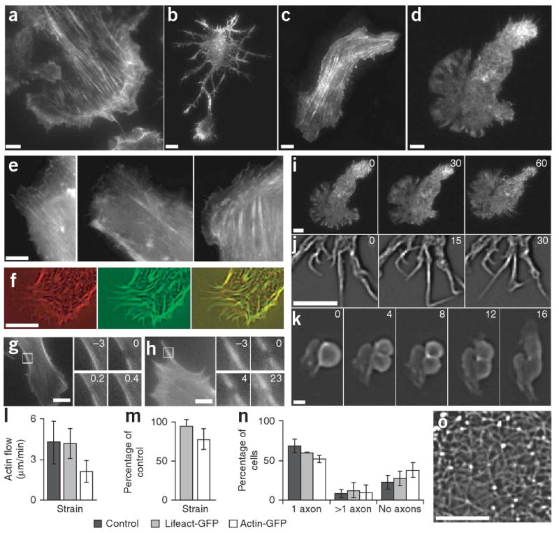
Characterization of Lifeact-GFP in vivo. (a–j) TIRF microscopy images of Lifeact-GFP transiently expressed in mouse embryonic fibroblasts (a), primary rat hippocampal neurons (b and time series of filopodial dynamics in j), MDCK cells (c) and mouse dendritic cells (d and a time series of chemotaxis in i). Epifluorescence images of actin-GFP (left), Lifeact-GFP (middle) and GFP-utrophin (right) in fibroblasts (e). MDCK cells transfected with actin-RFP and Lifeact-GFP, and imaged by widefield microscopy (RFP, left; GFP, middle; overlay, right; f). Fluorescence recovery after photobleaching in fibroblasts transfected with actin-GFP (g) and Lifeact-GFP (h), where numbers in insets (magnification of the boxed areas) indicate time relative to bleaching in seconds. (k) Time series (in minutes) of an MDCK cell undergoing cytokinesis, showing Lifeact-GFP staining in the contractile ring. (l) Velocity of lamellipodial retrograde actin flow in untransfected (control) or transiently transfected (Lifeact-GFP or actin-GFP) fibroblasts measured from kymograph traces. (m) Chemotactic speed of transiently transfected dendritic cells relative to untransfected cells. (n) Quantification of neuronal polarization 3 d after transient transfection. Data are averages ± s.d. from at least three experiments. (o) Cortical actin network of a hippocampal neuron transfected with Lifeact-GFP. Scale bars, 5 μm except 1 μm in j.
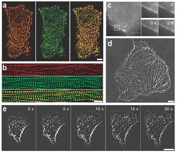
F-Lifeact staining in fixed and living samples. (a,b) MDCK cells (a) and cryo-sections of mouse skeletal muscle (b) fixed with paraformaldehyde and stained with F-Lifeact (green) and phalloidin-Cy3 (red, overlay in yellow). (c) Fluorescence recovery after photobleaching of a paraformaldehyde-fixed F-Lifeact–stained mouse embryonic fibroblast. Numbers in insets (magnification of the boxed areas) indicate time relative to bleaching in seconds. (d,e) Mouse embryonic fibroblasts (d) and human primary neutrophil granulocytes (e) were scrape-loaded with F-Lifeact, replated and cell spreading was visualized with TIRF microscopy. Scale bars, 5 μm.
Comment in
-
Lifeact cannot visualize some forms of stress-induced twisted F-actin.Nat Methods. 2009 May;6(5):317. doi: 10.1038/nmeth0509-317. Nat Methods. 2009. PMID: 19404250 No abstract available.
References
Publication types
MeSH terms
Substances
Grants and funding
LinkOut - more resources
Full Text Sources
Other Literature Sources
Molecular Biology Databases
Research Materials
