. 2008 Sep;155(1):62-72.
doi: 10.1038/bjp.2008.231. Epub 2008 Jun 9.

Bimodal effects of the Kv7 channel activator retigabine on vascular K+ currents

Affiliations

Bimodal effects of the Kv7 channel activator retigabine on vascular K+ currents

Sym Yeung et al. Br J Pharmacol. 2008 Sep.

Abstract

Background and purpose: This study investigated the functional and electrophysiological effects of the Kv7 channel activator, retigabine, on murine portal vein smooth muscle.

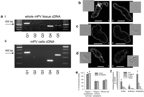
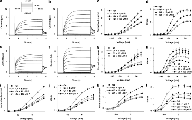
Experimental approach: KCNQ gene expression was determined by reverse transcriptase polymerase chain reaction (RT-PCR) and immunocytochemical experiments. Whole cell voltage clamp and current clamp were performed on isolated myocytes from murine portal vein. Isometric tension recordings were performed on whole portal veins. K+ currents generated by KCNQ4 and KCNQ5 expression were recorded by two-electrode voltage clamp in Xenopus oocytes.

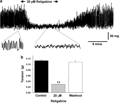
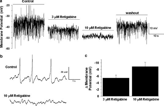
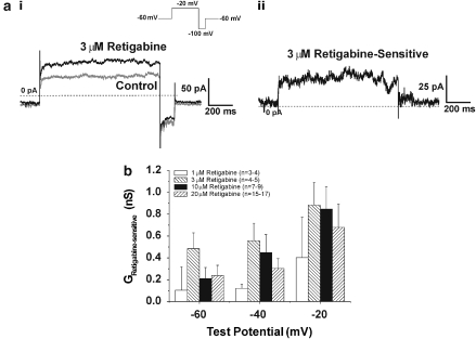
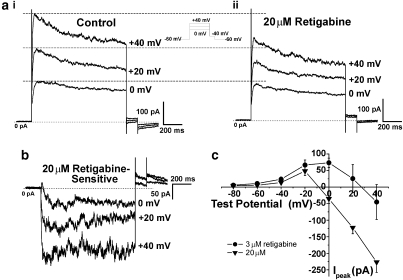
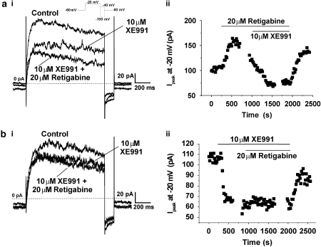
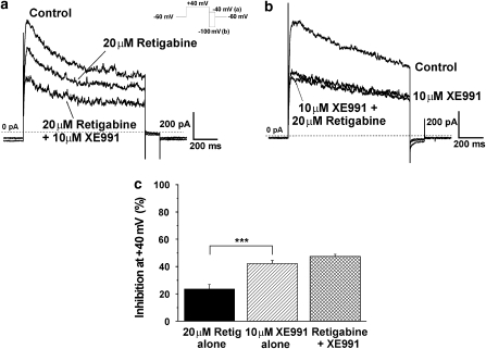
Key results: KCNQ1, 4 and 5 were expressed in mRNA derived from murine portal vein, either as whole tissue or isolated myocytes. Kv7.1 and Kv7.4 proteins were identified in the cell membranes of myocytes by immunocytochemistry. Retigabine (2-20 microM) suppressed spontaneous contractions in whole portal veins, hyperpolarized the membrane potential and augmented potassium currents at -20 mV. At more depolarized potentials, retigabine and flupirtine, decreased potassium currents. Both effects of retigabine were prevented by prior application of the K(v)7 blocker XE991 (10 muM). Recombinant KCNQ 4 or 5 channels were only activated by retigabine or flupirtine.

Conclusions and implications: The Kv7 channel activators retigabine and flupirtine have bimodal effects on vascular potassium currents, which are not seen with recombinant KCNQ channels. These results provide support for KCNQ4- or KCNQ5-encoded channels having an important functional impact in the vasculature.


Figures

Figure 1
Figure 1
Retigabine inhibits excitability of whole mPV tissue. (a) Example of isometric tension recording in the absence and presence of 20 μM retigabine. Lower traces show magnifications of the sections highlighted. (b) Bar chart shows mean tension in the absence (solid bars) and presence of retigabine (hatched bars) and upon washout (open bars). ** denotes statistical significance retigabine versus control at P<0.01.
Figure 2
Figure 2
Effect of retigabine on membrane potential recordings in current clamp mode. (a) Example of membrane potential in the absence and presence of retigabine. Each panel shows a 20 s sample of membrane potential recording either in the absence of retigabine or after 5 min application of 3 μM and then 10 μM retigabine. Right panel shows the membrane potential 7 min after washout of retigabine. Upward and downward deflections denote spontaneous depolarizations and hyperpolarizations. (b) shows an amplified view of membrane potential recordings in the absence and presence of 10 μM retigabine showing the lack of spontaneous membrane depolarizations in the presence of this agent. Panel (c) shows the mean membrane hyperpolarization produced by 3 and 10 μM retigabine (n=5 and 6, respectively).
Figure 3
Figure 3
Activation of K+ currents by different concentrations of retigabine. Panel (ai) shows currents evoked by stepping from −60 to −20 mV in the absence and presence of 3 μM retigabine. (aii) shows the retigabine-sensitive current from (ai). (b) shows the effect of a range of retigabine concentrations on K+ conductance (I/V-Vr) at −60, −40 and −20 mV.
Figure 4
Figure 4
Retigabine inhibited the K+ currents at positive potentials in a concentration-dependent manner. Current recordings at 0, +20 and +40 mV in the (ai) absence and (aii) presence of 20 μM retigabine. (b) Currents sensitive to retigabine at these potentials. (c) Current–voltage plot of peak current amplitudes sensitive to 3 and 20 μM retigabine.
Figure 5
Figure 5
The effects of retigabine were sensitive to XE991. Panel (ai) show currents at −20 mV in the presence of retigabine and the subsequent application of XE991. (aii) shows the amplitude of currents at −20 mV over time for the experiment in (ai). (bi) shows the lack of effect of retigabine on currents at −20 mV in a cell bathed in 10 μM XE991. (bii) shows the amplitude of currents at −20 mV over time for the experiment in (bi).
Figure 6
Figure 6
The inhibitory effects of retigabine and XE991 were not additive. Current recordings at VT +40 mV with (a) retigabine alone followed by retigabine with added XE991. The inhibition by 10 μM XE991 alone (b) was not altered when retigabine was added. (c) Histogram shows mean±s.e.m. (all n=7) block of mPV current (***P<0.001).
Figure 7
Figure 7
Expression of mRNA and protein in mPV tissue and cells. (a) RT-PCR analysis of KCNQ genes in RNA extracted from mPV tissue (i) or a pool of 20 isolated myocytes (ii). In panel 7aii, the size of the products are different from those in 7ai because a nested PCR was used. Immunocytochemical staining of murine portal vein myocytes for (b) Kv7.1 and (c and d) Kv7.4. The right-hand panels show control experiments in the presence of an antigenic peptide (b and d) or in the absence of primary antibodies (c). White circles in panel bi indicate Regions 1 and 2, which were used to analyse the localization of fluorescence. A dotted line was used where necessary to outline the contour of a cell, due to its low fluorescence. The insets show transmitted light images of respective cells. Calibration: 10 μm. (e) Summarized data on localization of Kv7 fluorescence in the cells showing the percentage of fluorescent pixels in the region within ∼1 μm of plasma membrane (Region 1), in the deep cytoplasm (Region 2) and in the whole confocal plane. (f) Summary data showing the specificity of labelling, confirmed by a significant decrease in fluorescence either after preincubation with antigenic peptide (AgP) or in the absence of primary antibodies. 1′=Region1; 2′=Region 2 *Significantly different from other values shown. #: antigenic peptide not available.
Figure 8
Figure 8
Activation of heterologously expressed KCNQ4 and KCNQ5 by retigabine and flupirtine. Typical current traces of oocytes expressing KCNQ4 (a) and KCNQ5 (e) before and after (KCNQ4 (b); KCNQ5 (f)) the application of 10 μM retigabine (R). The voltage protocol used for these experiments is shown as inset in panel a. I/V curves of KCNQ4 (c and i) and KCNQ5 (g and k) as a summary of the current recordings, which were obtained from oocytes measured with different retigabine (KCNQ4 (c) and KCNQ5 (g)) and different flupirtine (F) concentrations (KCNQ4 (i) and KCNQ5 (k)). I/Imax curves of KCNQ4 and KCNQ5 as a function of voltage obtained from tail current analysis and different retigabine (KCNQ4 (d) and KCNQ5 (h)) or flupirtine (KCNQ4 (j) and KCNQ5 (l)) concentrations. For all experiments n=8.

Similar articles

Cited by

References

    1. Alexander SPH, Mathie A, Peters JA. Guide to Receptors and Channels (GRAC), 3rd edn. Br J Pharmacol. 2008;153 (Suppl. 2):S1–S209. - PMC - PubMed
    1. Brueggemann LI, Moran CJ, Barakat JA, Yehy JZ, Cribbs LL, Byron KL. Vasopressin stimulates action potential firing by protein kinase C dependent inhibition of KCNQ5 in A7r5 rat aortic smooth muscle cells. Am J Physiol-Heart. 2006;292:H1352–H1363. - PMC - PubMed
    1. Dupuis DS, Olesen S-P, Jespersen T, Christensen JK, Christophersen P, Jensen BS. Activation of KCNQ5 Channels stably expressed in HEK293 cells by BMS-204352. Eur J Pharmacol. 2002;437:129–137. - PubMed
    1. Grunnet M, Jespersen T, Rasmussen HB, Ljungstrom T, Jorgensen NK, Olesen SP, et al. KCNE4 is an inhibitory subunit to the KCNQ1 channel. J Physiol. 2002;542:119–130. - PMC - PubMed
    1. Jentsch TJ. Neuronal KCNQ potassium channels: physiology and role in disease. Nat Rev Neurosci. 2000;1:21–30. - PubMed

Publication types

MeSH terms